6HUR | pdb_00006hur

2'-fucosyllactose and 3-fucosyllactose binding protein from Bifidobacterium longum infantis, bound with 2'-fucosyllactose


Experimental Data Snapshot

  • Method: X-RAY DIFFRACTION
  • Resolution: 1.30 Å
  • R-Value Free: 
    0.171 (Depositor), 0.171 (DCC) 
  • R-Value Work: 
    0.151 (Depositor), 0.153 (DCC) 

wwPDB Validation 3D Report Full Report

Validation slider image for 6HUR

Ligand Structure Quality Assessment 


This is version 2.1 of the entry. See complete history

Literature

Evolutionary adaptation in fucosyllactose uptake systems supports bifidobacteria-infant symbiosis.

Sakanaka, M.Hansen, M.E.Gotoh, A.Katoh, T.Yoshida, K.Odamaki, T.Yachi, H.Sugiyama, Y.Kurihara, S.Hirose, J.Urashima, T.Xiao, J.Z.Kitaoka, M.Fukiya, S.Yokota, A.Lo Leggio, L.Abou Hachem, M.Katayama, T.

(2019) Sci Adv 5: eaaw7696-eaaw7696

  • DOI: https://doi.org/10.1126/sciadv.aaw7696
  • Primary Citation Related Structures: 
    6HUR, 6HUS

  • PubMed Abstract: 

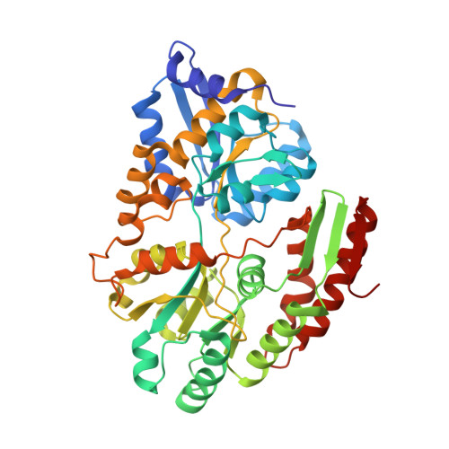
    The human gut microbiota established during infancy has persistent effects on health. In vitro studies have suggested that human milk oligosaccharides (HMOs) in breast milk promote the formation of a bifidobacteria-rich microbiota in infant guts; however, the underlying molecular mechanism remains elusive. Here, we characterized two functionally distinct but overlapping fucosyllactose transporters (FL transporter-1 and -2) from Bifidobacterium longum subspecies infantis . Fecal DNA and HMO consumption analyses, combined with deposited metagenome data mining, revealed that FL transporter-2 is primarily associated with the bifidobacteria-rich microbiota formation in breast-fed infant guts. Structural analyses of the solute-binding protein (SBP) of FL transporter-2 complexed with 2'-fucosyllactose and 3-fucosyllactose, together with phylogenetic analysis of SBP homologs of both FL transporters, highlight a unique adaptation strategy of Bifidobacterium to HMOs, in which the gain-of-function mutations enable FL transporter-2 to efficiently capture major fucosylated HMOs. Our results provide a molecular insight into HMO-mediated symbiosis and coevolution between bifidobacteria and humans.


  • Organizational Affiliation
    • Faculty of Bioresources and Environmental Sciences, Ishikawa Prefectural University, Nonoichi, Ishikawa 921-8836, Japan.

Macromolecule Content 

  • Total Structure Weight: 49.63 kDa 
  • Atom Count: 3,992 
  • Modeled Residue Count: 428 
  • Deposited Residue Count: 428 
  • Unique protein chains: 1

Macromolecules

Find similar proteins by:|  3D Structure
Entity ID: 1
MoleculeChains  Sequence LengthOrganismDetailsImage
ABC transporter substrate-binding protein428Bifidobacterium longum subsp. infantisMutation(s): 0 
Gene Names: BFS25_04965
UniProt
Find proteins for A0A1S2VYK0 (Bifidobacterium longum subsp. infantis)
Explore A0A1S2VYK0 
Go to UniProtKB:  A0A1S2VYK0
Entity Groups
Sequence Clusters30% Identity50% Identity70% Identity90% Identity95% Identity100% Identity
UniProt GroupA0A1S2VYK0
Sequence Annotations
Expand
Reference Sequence

Oligosaccharides

Help  
Entity ID: 2
MoleculeChains Length2D Diagram GlycosylationD Interactions
alpha-L-fucopyranose-(1-2)-beta-D-galactopyranose-(1-4)-alpha-D-glucopyranose
B
3N/A
Glycosylation Resources
GlyTouCan: G94133JU
GlyCosmos: G94133JU
GlyGen: G94133JU

Small Molecules

Ligands 4 Unique
IDChains Name / Formula / InChI Key2D Diagram3D Interactions
MES

Query on MES


Download:Ideal Coordinates CCD File
J [auth A],
K [auth A]
2-(N-MORPHOLINO)-ETHANESULFONIC ACID
C6 H13 N O4 S
SXGZJKUKBWWHRA-UHFFFAOYSA-N
BGC

Query on BGC


Download:Ideal Coordinates CCD File
D [auth A]beta-D-glucopyranose
C6 H12 O6
WQZGKKKJIJFFOK-VFUOTHLCSA-N
PG0

Query on PG0


Download:Ideal Coordinates CCD File
C [auth A]2-(2-METHOXYETHOXY)ETHANOL
C5 H12 O3
SBASXUCJHJRPEV-UHFFFAOYSA-N
ZN

Query on ZN


Download:Ideal Coordinates CCD File
E [auth A],
F [auth A],
G [auth A],
H [auth A],
I [auth A]
ZINC ION
Zn
PTFCDOFLOPIGGS-UHFFFAOYSA-N

Experimental Data & Validation

Experimental Data

  • Method: X-RAY DIFFRACTION
  • Resolution: 1.30 Å
  • R-Value Free:  0.171 (Depositor), 0.171 (DCC) 
  • R-Value Work:  0.151 (Depositor), 0.153 (DCC) 
Space Group: H 3
Unit Cell:
Length ( Å )Angle ( ˚ )
a = 134α = 90
b = 134β = 90
c = 59.7γ = 120
Software Package:
Software NamePurpose
PHENIXrefinement
PDB_EXTRACTdata extraction
XDSdata reduction
XSCALEdata scaling
Cootmodel building
PHENIXmodel building
PHENIXphasing

Structure Validation

View Full Validation Report


Ligand Structure Quality Assessment 


Entry History 

& Funding Information

Deposition Data


Funding OrganizationLocationGrant Number
Danish Council for Independent ResearchDenmarkDFF-4002-00297

Revision History  (Full details and data files)

  • Version 1.0: 2019-09-04
    Type: Initial release
  • Version 1.1: 2019-09-18
    Changes: Data collection, Database references
  • Version 2.0: 2020-07-29
    Type: Remediation
    Reason: Carbohydrate remediation
    Changes: Advisory, Atomic model, Data collection, Derived calculations, Structure summary
  • Version 2.1: 2024-05-15
    Changes: Data collection, Database references, Derived calculations, Structure summary