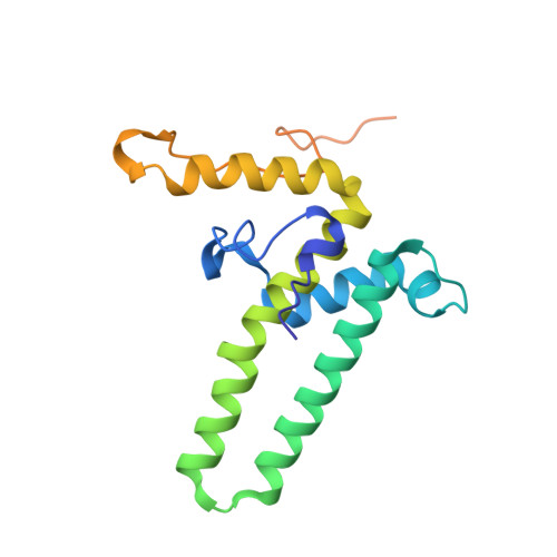
Structure of Mutant Hepatitis B Core Protein Capsids with Premature Secretion Phenotype.
Bottcher, B., Nassal, M.(2018) J Mol Biology 430: 4941-4954
- PubMed: 30539760
- DOI: https://doi.org/10.1016/j.jmb.2018.10.018
- Primary Citation of Related Structures:
6HTX, 6HU4, 6HU7 - PubMed Abstract:
Hepatitis B virus is a major human pathogen that consists of a viral genome surrounded by an icosahedrally ordered core protein and a polymorphic, lipidic envelope that is densely packed with surface proteins. A point mutation in the core protein in which a phenylalanine at position 97 is exchanged for a smaller leucine leads to premature envelopment of the capsid before the genome maturation is fully completed. We have used electron cryo-microscopy and image processing to investigate how the point mutation affects the structure of the capsid at 2.6- to 2.8 Å-resolution. We found that in the mutant the smaller side chain at position 97 is displaced, increasing the size of an adjacent pocket in the center of the spikes of the capsid. In the mutant, this pocket is filled with an unknown density. Phosphorylation of serine residues in the unresolved C-terminal domain of the mutant leaves the structure of the ordered capsid largely unchanged. However, we were able to resolve several previously unresolved residues downstream of proline 144 that precede the phosphorylation-sites. These residues pack against the neighboring subunits and increase the inter-dimer contact suggesting that the C-termini play an important role in capsid stabilization and provide a much larger interaction interface than previously observed.
- Department of Biochemistry, Cryo Electron Microscopy, Julius Maximillian's University, Josef-Schneider Straße 2, 97080 Würzburg, Germany. Electronic address: bettina.boettcher@uni-wuerzburg.de.
Organizational Affiliation:


