6HRO | pdb_00006hro

Crystal structure of Ebolavirus glycoprotein in complex with inhibitor 118a


Experimental Data Snapshot

  • Method: X-RAY DIFFRACTION
  • Resolution: 2.30 Å
  • R-Value Free: 
    0.212 (Depositor), 0.222 (DCC) 
  • R-Value Work: 
    0.180 (Depositor), 0.191 (DCC) 
  • R-Value Observed: 
    0.182 (Depositor) 

wwPDB Validation 3D Report Full Report

Validation slider image for 6HRO

Ligand Structure Quality Assessment 


This is version 2.1 of the entry. See complete history

Literature

Structure-Based in Silico Screening Identifies a Potent Ebolavirus Inhibitor from a Traditional Chinese Medicine Library.

Shaikh, F.Zhao, Y.Alvarez, L.Iliopoulou, M.Lohans, C.Schofield, C.J.Padilla-Parra, S.Siu, S.W.I.Fry, E.E.Ren, J.Stuart, D.I.

(2019) J Med Chem 62: 2928-2937

  • DOI: https://doi.org/10.1021/acs.jmedchem.8b01328
  • Primary Citation Related Structures: 
    6HRO, 6HS4

  • PubMed Abstract: 

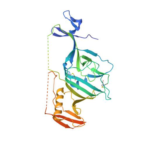
    Potent Ebolavirus (EBOV) inhibitors will help to curtail outbreaks such as that which occurred in 2014-16 in West Africa. EBOV has on its surface a single glycoprotein (GP) critical for viral entry and membrane fusion. Recent high-resolution complexes of EBOV GP with a variety of approved drugs revealed that binding to a common cavity prevented fusion of the virus and endosomal membranes, inhibiting virus infection. We performed docking experiments, screening a database of natural compounds to identify those likely to bind at this site. Using both inhibition assays of HIV-1-derived pseudovirus cell entry and structural analyses of the complexes of the compounds with GP, we show here that two of these compounds attach in the common binding cavity, out of eight tested. In both cases, two molecules bind in the cavity. The two compounds are chemically similar, but the tighter binder has an additional chlorine atom that forms good halogen bonds to the protein and achieves an IC 50 of 50 nM, making it the most potent GP-binding EBOV inhibitor yet identified, validating our screening approach for the discovery of novel antiviral compounds.


  • Organizational Affiliation
    • Department of Computer and Information Science, Faculty of Science and Technology , University of Macau , E11, Macau 999078 , China.

Macromolecule Content 

  • Total Structure Weight: 58.69 kDa 
  • Atom Count: 3,390 
  • Modeled Residue Count: 388 
  • Deposited Residue Count: 499 
  • Unique protein chains: 2

Macromolecules

Find similar proteins by:|  3D Structure
Entity ID: 1
MoleculeChains  Sequence LengthOrganismDetailsImage
Envelope glycoprotein,Envelope glycoprotein,Envelope glycoprotein331Ebola virus - Mayinga, Zaire, 1976Mutation(s): 1 
Gene Names: GP
UniProt
Find proteins for Q05320 (Zaire ebolavirus (strain Mayinga-76))
Explore Q05320 
Go to UniProtKB:  Q05320
Entity Groups
Sequence Clusters30% Identity50% Identity70% Identity90% Identity95% Identity100% Identity
UniProt GroupQ05320
Glycosylation
Glycosylation Sites: 4
Sequence Annotations
Expand
Reference Sequence
Find similar proteins by:|  3D Structure
Entity ID: 2
MoleculeChains  Sequence LengthOrganismDetailsImage
Envelope glycoprotein168Ebola virus - Mayinga, Zaire, 1976Mutation(s): 1 
Gene Names: GP
UniProt
Find proteins for Q05320 (Zaire ebolavirus (strain Mayinga-76))
Explore Q05320 
Go to UniProtKB:  Q05320
Entity Groups
Sequence Clusters30% Identity50% Identity70% Identity90% Identity95% Identity100% Identity
UniProt GroupQ05320
Glycosylation
Glycosylation Sites: 2
Sequence Annotations
Expand
Reference Sequence

Oligosaccharides

Help  
Entity ID: 3
MoleculeChains Length2D Diagram GlycosylationD Interactions
alpha-D-mannopyranose-(1-3)-[alpha-D-mannopyranose-(1-6)]beta-D-mannopyranose-(1-4)-2-acetamido-2-deoxy-beta-D-glucopyranose-(1-4)-2-acetamido-2-deoxy-beta-D-glucopyranose
C
5N-Glycosylation
Glycosylation Resources
GlyTouCan: G22768VO
GlyCosmos: G22768VO
GlyGen: G22768VO

Small Molecules

Ligands 4 Unique
IDChains Name / Formula / InChI Key2D Diagram3D Interactions
GKZ

Query on GKZ


Download:Ideal Coordinates CCD File
J [auth A],
O [auth B]
1-[2-[4-[4-(4-chlorophenyl)-3-methyl-1~{H}-pyrazol-5-yl]-3-oxidanyl-phenoxy]ethyl]piperidin-1-ium-4-carboxamide
C24 H28 Cl N4 O3
FIAWYOJOIHFYFE-UHFFFAOYSA-O
NAG

Query on NAG


Download:Ideal Coordinates CCD File
D [auth A],
E [auth A],
F [auth A],
G [auth A],
L [auth B]
2-acetamido-2-deoxy-beta-D-glucopyranose
C8 H15 N O6
OVRNDRQMDRJTHS-FMDGEEDCSA-N
GOL

Query on GOL


Download:Ideal Coordinates CCD File
H [auth A],
I [auth A],
M [auth B],
N [auth B]
GLYCEROL
C3 H8 O3
PEDCQBHIVMGVHV-UHFFFAOYSA-N
DMS

Query on DMS


Download:Ideal Coordinates CCD File
K [auth A]DIMETHYL SULFOXIDE
C2 H6 O S
IAZDPXIOMUYVGZ-UHFFFAOYSA-N

Experimental Data & Validation

Experimental Data

  • Method: X-RAY DIFFRACTION
  • Resolution: 2.30 Å
  • R-Value Free:  0.212 (Depositor), 0.222 (DCC) 
  • R-Value Work:  0.180 (Depositor), 0.191 (DCC) 
  • R-Value Observed: 0.182 (Depositor) 
Space Group: H 3 2
Unit Cell:
Length ( Å )Angle ( ˚ )
a = 115.12α = 90
b = 115.12β = 90
c = 308.86γ = 120
Software Package:
Software NamePurpose
PHENIXrefinement
xia2data reduction
xia2data scaling
REFMACphasing

Structure Validation

View Full Validation Report


Ligand Structure Quality Assessment 


Entry History 

& Funding Information

Deposition Data


Funding OrganizationLocationGrant Number
United Kingdom--

Revision History  (Full details and data files)

  • Version 1.0: 2019-02-27
    Type: Initial release
  • Version 1.1: 2019-03-06
    Changes: Data collection, Structure summary
  • Version 1.2: 2019-04-10
    Changes: Data collection, Database references
  • Version 2.0: 2020-07-29
    Type: Remediation
    Reason: Carbohydrate remediation
    Changes: Atomic model, Data collection, Derived calculations, Structure summary
  • Version 2.1: 2024-10-16
    Changes: Data collection, Database references, Derived calculations, Structure summary