6HLX | pdb_00006hlx

Structure of the PBP AgaA in complex with agropinic acid from A.tumefacien R10


Experimental Data Snapshot

  • Method: X-RAY DIFFRACTION
  • Resolution: 1.65 Å
  • R-Value Free: 
    0.174 (Depositor), 0.164 (DCC) 
  • R-Value Work: 
    0.156 (Depositor) 
  • R-Value Observed: 
    0.157 (Depositor) 

Starting Models: experimental
View more details

wwPDB Validation 3D Report Full Report

Validation slider image for 6HLX

Ligand Structure Quality Assessment 


This is version 1.2 of the entry. See complete history

Literature

Structural basis for two efficient modes of agropinic acid opine import into the bacterial pathogenAgrobacterium tumefaciens.

Marty, L.Vigouroux, A.Aumont-Nicaise, M.Pelissier, F.Meyer, T.Lavire, C.Dessaux, Y.Morera, S.

(2019) Biochem J 476: 165-178

  • DOI: https://doi.org/10.1042/BCJ20180861
  • Primary Citation Related Structures: 
    6HLX, 6HLY, 6HLZ, 6HM2

  • PubMed Abstract: 

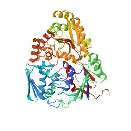
    Agrobacterium tumefaciens pathogens genetically modify their host plants to drive the synthesis of opines in plant tumors. The mannityl-opine family encompasses mannopine, mannopinic acid, agropine and agropinic acid. These opines serve as nutrients and are imported into bacteria via periplasmic-binding proteins (PBPs) in association with ABC transporters. Structural and affinity data on agropine and agropinic acid opines bound to PBPs are currently lacking. Here, we investigated the molecular basis of AgtB and AgaA, proposed as the specific PBP for agropine and agropinic acid import, respectively. Using genetic approaches and affinity measurements, we identified AgtB and its transporter as responsible for agropine uptake in agropine-assimilating agrobacteria. Nonetheless, we showed that AgtB binds agropinic acid with a higher affinity than agropine, and we structurally characterized the agropinic acid-binding mode through three crystal structures at 1.4, 1.74 and 1.9 Å resolution. In the crystallization time course, obtaining a crystal structure of AgtB with agropine was unsuccessful due to the spontaneous lactamization of agropine into agropinic acid. AgaA binds agropinic acid only with a similar affinity in nanomolar range as AgtB. The structure of AgaA bound to agropinic acid at 1.65 Å resolution defines a different agropinic acid-binding signature. Our work highlights the structural and functional characteristics of two efficient agropinic acid assimilation pathways, of which one is also involved in agropine assimilation.


  • Organizational Affiliation
    • Institute for Integrative Biology of the Cell (I2BC), CNRS CEA Univ. Paris-Sud, Université Paris-Saclay, Avenue de la Terrasse, 91198 Gif-sur-Yvette, France.

Macromolecule Content 

  • Total Structure Weight: 57.39 kDa 
  • Atom Count: 4,502 
  • Modeled Residue Count: 491 
  • Deposited Residue Count: 492 
  • Unique protein chains: 1

Macromolecules

Find similar proteins by:|  3D Structure
Entity ID: 1
MoleculeChains  Sequence LengthOrganismDetailsImage
AgaA492Agrobacterium tumefaciensMutation(s): 0 
Gene Names: agaAA6U90_18825At1D1609_52290
UniProt
Find proteins for O50260 (Agrobacterium tumefaciens)
Explore O50260 
Go to UniProtKB:  O50260
Entity Groups
Sequence Clusters30% Identity50% Identity70% Identity90% Identity95% Identity100% Identity
UniProt GroupO50260
Sequence Annotations
Expand
Reference Sequence

Small Molecules

Ligands 8 Unique
IDChains Name / Formula / InChI Key2D Diagram3D Interactions
G9Z

Query on G9Z


Download:Ideal Coordinates CCD File
E [auth A]agropinic acid
C11 H17 N O8
WYIHPABVKQQJFB-SZWOQXJISA-N
MES

Query on MES


Download:Ideal Coordinates CCD File
F [auth A]2-(N-MORPHOLINO)-ETHANESULFONIC ACID
C6 H13 N O4 S
SXGZJKUKBWWHRA-UHFFFAOYSA-N
PEG

Query on PEG


Download:Ideal Coordinates CCD File
LA [auth A]DI(HYDROXYETHYL)ETHER
C4 H10 O3
MTHSVFCYNBDYFN-UHFFFAOYSA-N
SO4

Query on SO4


Download:Ideal Coordinates CCD File
T [auth A],
U [auth A],
V [auth A],
W [auth A]
SULFATE ION
O4 S
QAOWNCQODCNURD-UHFFFAOYSA-L
GOL

Query on GOL


Download:Ideal Coordinates CCD File
B [auth A],
C [auth A],
D [auth A]
GLYCEROL
C3 H8 O3
PEDCQBHIVMGVHV-UHFFFAOYSA-N
ZN

Query on ZN


Download:Ideal Coordinates CCD File
N [auth A]
O [auth A]
P [auth A]
Q [auth A]
R [auth A]
N [auth A],
O [auth A],
P [auth A],
Q [auth A],
R [auth A],
S [auth A]
ZINC ION
Zn
PTFCDOFLOPIGGS-UHFFFAOYSA-N
EDO

Query on EDO


Download:Ideal Coordinates CCD File
AA [auth A]
BA [auth A]
CA [auth A]
DA [auth A]
EA [auth A]
AA [auth A],
BA [auth A],
CA [auth A],
DA [auth A],
EA [auth A],
FA [auth A],
GA [auth A],
HA [auth A],
IA [auth A],
JA [auth A],
KA [auth A],
X [auth A],
Y [auth A],
Z [auth A]
1,2-ETHANEDIOL
C2 H6 O2
LYCAIKOWRPUZTN-UHFFFAOYSA-N
ACT

Query on ACT


Download:Ideal Coordinates CCD File
G [auth A]
H [auth A]
I [auth A]
J [auth A]
K [auth A]
G [auth A],
H [auth A],
I [auth A],
J [auth A],
K [auth A],
L [auth A],
M [auth A]
ACETATE ION
C2 H3 O2
QTBSBXVTEAMEQO-UHFFFAOYSA-M

Experimental Data & Validation

Experimental Data

  • Method: X-RAY DIFFRACTION
  • Resolution: 1.65 Å
  • R-Value Free:  0.174 (Depositor), 0.164 (DCC) 
  • R-Value Work:  0.156 (Depositor) 
  • R-Value Observed: 0.157 (Depositor) 
Space Group: P 65 2 2
Unit Cell:
Length ( Å )Angle ( ˚ )
a = 89.26α = 90
b = 89.26β = 90
c = 285.85γ = 120
Software Package:
Software NamePurpose
BUSTERrefinement
XDSdata reduction
XSCALEdata scaling
PHASERphasing

Structure Validation

View Full Validation Report


Ligand Structure Quality Assessment 


Entry History 

Deposition Data

Revision History  (Full details and data files)

  • Version 1.0: 2018-12-26
    Type: Initial release
  • Version 1.1: 2019-01-23
    Changes: Data collection, Database references
  • Version 1.2: 2024-01-24
    Changes: Data collection, Database references, Derived calculations, Refinement description