6HJX | pdb_00006hjx

X-ray structure of a pentameric ligand gated ion channel from Erwinia chrysanthemi (ELIC) 7'C pore mutant (L238C) in complex with nanobody 72


Experimental Data Snapshot

  • Method: X-RAY DIFFRACTION
  • Resolution: 2.50 Å
  • R-Value Free: 
    0.245 (Depositor), 0.247 (DCC) 
  • R-Value Work: 
    0.204 (Depositor), 0.206 (DCC) 
  • R-Value Observed: 
    0.206 (Depositor) 

Starting Model: experimental
View more details

wwPDB Validation 3D Report Full Report

Validation slider image for 6HJX

Ligand Structure Quality Assessment 


This is version 1.4 of the entry. See complete history

Literature

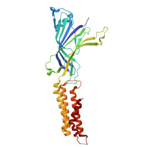
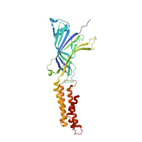
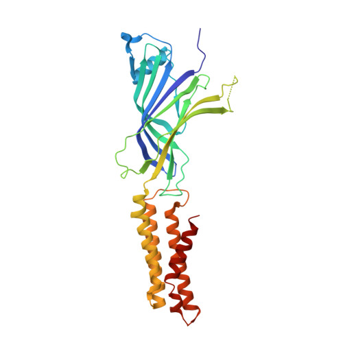
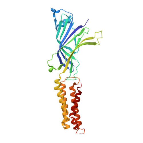
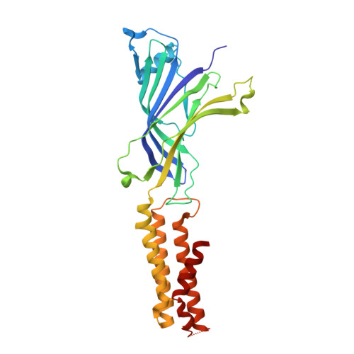
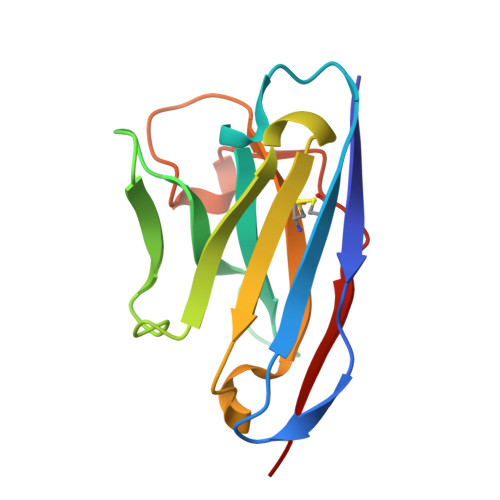
A lipid site shapes the agonist response of a pentameric ligand-gated ion channel.

Henault, C.M.Govaerts, C.Spurny, R.Brams, M.Estrada-Mondragon, A.Lynch, J.Bertrand, D.Pardon, E.Evans, G.L.Woods, K.Elberson, B.W.Cuello, L.G.Brannigan, G.Nury, H.Steyaert, J.Baenziger, J.E.Ulens, C.

(2019) Nat Chem Biol 15: 1156-1164

  • DOI: https://doi.org/10.1038/s41589-019-0369-4
  • Primary Citation Related Structures: 
    6HJX, 6HJY, 6HK0

  • PubMed Abstract: 

    Phospholipids are key components of cellular membranes and are emerging as important functional regulators of different membrane proteins, including pentameric ligand-gated ion channels (pLGICs). Here, we take advantage of the prokaryote channel ELIC (Erwinia ligand-gated ion channel) as a model to understand the determinants of phospholipid interactions in this family of receptors. A high-resolution structure of ELIC in a lipid-bound state reveals a phospholipid site at the lower half of pore-forming transmembrane helices M1 and M4 and at a nearby site for neurosteroids, cholesterol or general anesthetics. This site is shaped by an M4-helix kink and a Trp-Arg-Pro triad that is highly conserved in eukaryote GABA A/C and glycine receptors. A combined approach reveals that M4 is intrinsically flexible and that M4 deletions or disruptions of the lipid-binding site accelerate desensitization in ELIC, suggesting that lipid interactions shape the agonist response. Our data offer a structural context for understanding lipid modulation in pLGICs.


  • Organizational Affiliation
    • Department of Biochemistry, Microbiology and Immunology, University of Ottawa, Ottawa, Ontario, Canada.

Macromolecule Content 

  • Total Structure Weight: 249.24 kDa 
  • Atom Count: 17,226 
  • Modeled Residue Count: 2,131 
  • Deposited Residue Count: 2,164 
  • Unique protein chains: 6

Macromolecules

Find similar proteins by:|  3D Structure
Entity ID: 1
MoleculeChains  Sequence LengthOrganismDetailsImage
Cys-loop ligand-gated ion channel306Dickeya chrysanthemiMutation(s): 3 
Membrane Entity: Yes 
UniProt
Find proteins for P0C7B7 (Dickeya chrysanthemi)
Explore P0C7B7 
Go to UniProtKB:  P0C7B7
Entity Groups
Sequence Clusters30% Identity50% Identity70% Identity90% Identity95% Identity100% Identity
UniProt GroupP0C7B7
Sequence Annotations
Expand
Reference Sequence
Find similar proteins by:|  3D Structure
Entity ID: 2
MoleculeChains  Sequence LengthOrganismDetailsImage
Cys-loop ligand-gated ion channel309Dickeya chrysanthemiMutation(s): 3 
Membrane Entity: Yes 
UniProt
Find proteins for P0C7B7 (Dickeya chrysanthemi)
Explore P0C7B7 
Go to UniProtKB:  P0C7B7
Entity Groups
Sequence Clusters30% Identity50% Identity70% Identity90% Identity95% Identity100% Identity
UniProt GroupP0C7B7
Sequence Annotations
Expand
Reference Sequence
Find similar proteins by:|  3D Structure
Entity ID: 3
MoleculeChains  Sequence LengthOrganismDetailsImage
Cys-loop ligand-gated ion channel312Dickeya chrysanthemiMutation(s): 3 
Membrane Entity: Yes 
UniProt
Find proteins for P0C7B7 (Dickeya chrysanthemi)
Explore P0C7B7 
Go to UniProtKB:  P0C7B7
Entity Groups
Sequence Clusters30% Identity50% Identity70% Identity90% Identity95% Identity100% Identity
UniProt GroupP0C7B7
Sequence Annotations
Expand
Reference Sequence
Find similar proteins by:|  3D Structure
Entity ID: 4
MoleculeChains  Sequence LengthOrganismDetailsImage
Cys-loop ligand-gated ion channel310Dickeya chrysanthemiMutation(s): 3 
Membrane Entity: Yes 
UniProt
Find proteins for P0C7B7 (Dickeya chrysanthemi)
Explore P0C7B7 
Go to UniProtKB:  P0C7B7
Entity Groups
Sequence Clusters30% Identity50% Identity70% Identity90% Identity95% Identity100% Identity
UniProt GroupP0C7B7
Sequence Annotations
Expand
Reference Sequence
Find similar proteins by:|  3D Structure
Entity ID: 5
MoleculeChains  Sequence LengthOrganismDetailsImage
Cys-loop ligand-gated ion channel307Dickeya chrysanthemiMutation(s): 3 
Membrane Entity: Yes 
UniProt
Find proteins for P0C7B7 (Dickeya chrysanthemi)
Explore P0C7B7 
Go to UniProtKB:  P0C7B7
Entity Groups
Sequence Clusters30% Identity50% Identity70% Identity90% Identity95% Identity100% Identity
UniProt GroupP0C7B7
Sequence Annotations
Expand
Reference Sequence
Find similar proteins by:|  3D Structure
Entity ID: 6
MoleculeChains  Sequence LengthOrganismDetailsImage
nanobody 72
F, G, H, I, J
124Lama glamaMutation(s): 0 

Small Molecules

Ligands 5 Unique
IDChains Name / Formula / InChI Key2D Diagram3D Interactions
PTY
(Subject of Investigation/LOI)

Query on PTY


Download:Ideal Coordinates CCD File
L [auth A]PHOSPHATIDYLETHANOLAMINE
C40 H80 N O8 P
NJGIRBISCGPRPF-KXQOOQHDSA-N
LMT

Query on LMT


Download:Ideal Coordinates CCD File
M [auth A],
N [auth B],
P [auth C],
S [auth D],
U [auth E]
DODECYL-BETA-D-MALTOSIDE
C24 H46 O11
NLEBIOOXCVAHBD-QKMCSOCLSA-N
P6G

Query on P6G


Download:Ideal Coordinates CCD File
K [auth A],
O [auth B],
T [auth D],
V [auth E]
HEXAETHYLENE GLYCOL
C12 H26 O7
IIRDTKBZINWQAW-UHFFFAOYSA-N
MES

Query on MES


Download:Ideal Coordinates CCD File
Q [auth C]2-(N-MORPHOLINO)-ETHANESULFONIC ACID
C6 H13 N O4 S
SXGZJKUKBWWHRA-UHFFFAOYSA-N
NA

Query on NA


Download:Ideal Coordinates CCD File
R [auth C]SODIUM ION
Na
FKNQFGJONOIPTF-UHFFFAOYSA-N

Experimental Data & Validation

Experimental Data

  • Method: X-RAY DIFFRACTION
  • Resolution: 2.50 Å
  • R-Value Free:  0.245 (Depositor), 0.247 (DCC) 
  • R-Value Work:  0.204 (Depositor), 0.206 (DCC) 
  • R-Value Observed: 0.206 (Depositor) 
Space Group: P 1 21 1
Unit Cell:
Length ( Å )Angle ( ˚ )
a = 96.251α = 90
b = 156.277β = 102.88
c = 102.496γ = 90
Software Package:
Software NamePurpose
PHENIXrefinement
XDSdata reduction
Aimlessdata scaling
PHASERphasing

Structure Validation

View Full Validation Report


Ligand Structure Quality Assessment 


Entry History 

Deposition Data

Revision History  (Full details and data files)

  • Version 1.0: 2019-10-09
    Type: Initial release
  • Version 1.1: 2019-10-23
    Changes: Data collection, Database references
  • Version 1.2: 2019-11-27
    Changes: Database references
  • Version 1.3: 2024-01-17
    Changes: Data collection, Database references, Refinement description
  • Version 1.4: 2024-10-23
    Changes: Structure summary