6H9V | pdb_00006h9v

Crystal structure of deaminated P domain from norovirus strain Saga GII-4 in complex with Fuc


Experimental Data Snapshot

  • Method: X-RAY DIFFRACTION
  • Resolution: 1.52 Å
  • R-Value Free: 
    0.183 (Depositor), 0.183 (DCC) 
  • R-Value Work: 
    0.147 (Depositor), 0.149 (DCC) 
  • R-Value Observed: 
    0.149 (Depositor) 

wwPDB Validation 3D Report Full Report

Validation slider image for 6H9V

Ligand Structure Quality Assessment 


This is version 1.2 of the entry. See complete history

Literature

A post-translational modification of human Norovirus capsid protein attenuates glycan binding.

Mallagaray, A.Creutznacher, R.Dulfer, J.Mayer, P.H.O.Grimm, L.L.Orduna, J.M.Trabjerg, E.Stehle, T.Rand, K.D.Blaum, B.S.Uetrecht, C.Peters, T.

(2019) Nat Commun 10: 1320-1320

  • DOI: https://doi.org/10.1038/s41467-019-09251-5
  • Primary Citation Related Structures: 
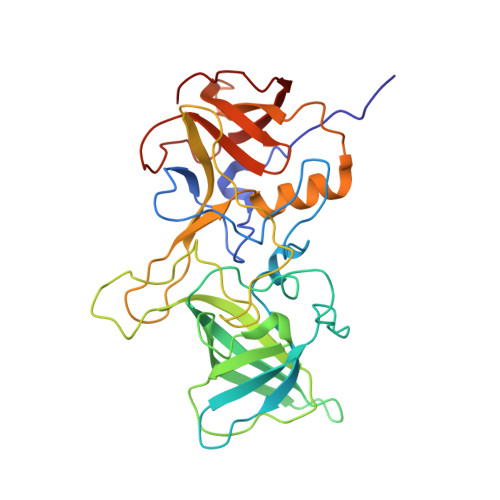
    6H9V

  • PubMed Abstract: 

    Attachment of human noroviruses to histo blood group antigens (HBGAs) is essential for infection, but how this binding event promotes the infection of host cells is unknown. Here, we employ protein NMR experiments supported by mass spectrometry and crystallography to study HBGA binding to the P-domain of a prevalent virus strain (GII.4). We report a highly selective transformation of asparagine 373, located in an antigenic loop adjoining the HBGA binding site, into an iso-aspartate residue. This spontaneous post-translational modification (PTM) proceeds with an estimated half-life of a few days at physiological temperatures, independent of the presence of HBGAs but dramatically affecting HBGA recognition. Sequence conservation and the surface-exposed position of this PTM suggest an important role in infection and immune recognition for many norovirus strains.


  • Organizational Affiliation
    • Institute of Chemistry and Metabolomics, Center of Structural and Cell Biology in Medicine (CSCM), University of Lübeck, Ratzeburger Allee 160, 23562, Lübeck, Germany.

Macromolecule Content 

  • Total Structure Weight: 68.49 kDa 
  • Atom Count: 5,656 
  • Modeled Residue Count: 612 
  • Deposited Residue Count: 618 
  • Unique protein chains: 1

Macromolecules

Find similar proteins by:|  3D Structure
Entity ID: 1
MoleculeChains  Sequence LengthOrganismDetailsImage
VP1
A, B
309Norovirus Hu/GII-4/Saga4/2006/JPMutation(s): 0 
UniProt
Find proteins for B5BTR7 (Norovirus Hu/GII-4/Saga4/2006/JP)
Explore B5BTR7 
Go to UniProtKB:  B5BTR7
Entity Groups
Sequence Clusters30% Identity50% Identity70% Identity90% Identity95% Identity100% Identity
UniProt GroupB5BTR7
Sequence Annotations
Expand
Reference Sequence

Experimental Data & Validation

Experimental Data

  • Method: X-RAY DIFFRACTION
  • Resolution: 1.52 Å
  • R-Value Free:  0.183 (Depositor), 0.183 (DCC) 
  • R-Value Work:  0.147 (Depositor), 0.149 (DCC) 
  • R-Value Observed: 0.149 (Depositor) 
Space Group: P 1 21 1
Unit Cell:
Length ( Å )Angle ( ˚ )
a = 54.29α = 90
b = 83.95β = 95.78
c = 65.13γ = 90
Software Package:
Software NamePurpose
PHENIXrefinement
XDSdata reduction
XSCALEdata scaling
REFMACphasing

Structure Validation

View Full Validation Report


Ligand Structure Quality Assessment 


Entry History 

& Funding Information

Deposition Data


Funding OrganizationLocationGrant Number
German Research FoundationGermanyBL1294/3-1

Revision History  (Full details and data files)

  • Version 1.0: 2019-04-03
    Type: Initial release
  • Version 1.1: 2020-07-29
    Type: Remediation
    Reason: Carbohydrate remediation
    Changes: Data collection, Derived calculations, Structure summary
  • Version 1.2: 2024-11-13
    Changes: Data collection, Database references, Structure summary