6H5X | pdb_00006h5x

Crystal structure of human Angiotensin-1 converting enzyme N-domain in complex with Omapatrilat.


Experimental Data Snapshot

  • Method: X-RAY DIFFRACTION
  • Resolution: 1.80 Å
  • R-Value Free: 
    0.204 (Depositor), 0.203 (DCC) 
  • R-Value Work: 
    0.164 (Depositor), 0.165 (DCC) 
  • R-Value Observed: 
    0.165 (Depositor) 

Starting Model: experimental
View more details

wwPDB Validation 3D Report Full Report

Validation slider image for 6H5X

Ligand Structure Quality Assessment 


This is version 2.2 of the entry. See complete history

Literature

Molecular Basis for Multiple Omapatrilat Binding Sites within the ACE C-Domain: Implications for Drug Design.

Cozier, G.E.Arendse, L.B.Schwager, S.L.Sturrock, E.D.Acharya, K.R.

(2018) J Med Chem 61: 10141-10154

  • DOI: https://doi.org/10.1021/acs.jmedchem.8b01309
  • Primary Citation Related Structures: 
    6H5W, 6H5X

  • PubMed Abstract: 

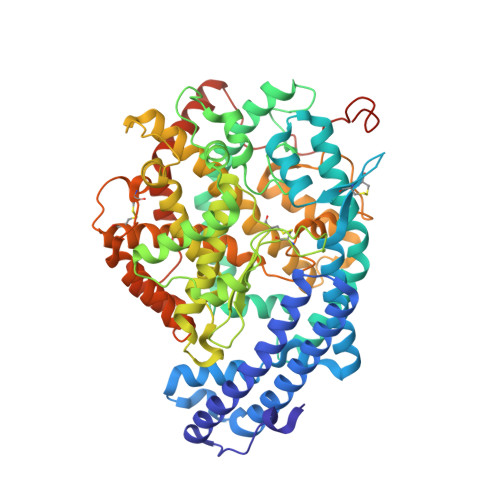
    Omapatrilat was designed as a vasopeptidase inhibitor with dual activity against the zinc metallopeptidases angiotensin-1 converting enzyme (ACE) and neprilysin (NEP). ACE has two homologous catalytic domains (nACE and cACE), which exhibit different substrate specificities. Here, we report high-resolution crystal structures of omapatrilat in complex with nACE and cACE and show omapatrilat has subnanomolar affinity for both domains. The structures show nearly identical binding interactions for omapatrilat in each domain, explaining the lack of domain selectivity. The cACE complex structure revealed an omapatrilat dimer occupying the cavity beyond the S 2 subsite, and this dimer had low micromolar inhibition of nACE and cACE. These results highlight residues beyond the S 2 subsite that could be exploited for domain selective inhibition. In addition, it suggests the possibility of either domain specific allosteric inhibitors that bind exclusively to the nonprime cavity or the potential for targeting specific substrates rather than completely inhibiting the enzyme.


  • Organizational Affiliation
    • Department of Biology and Biochemistry , University of Bath , Claverton Down , Bath BA2 7AY , United Kingdom.

Macromolecule Content 

  • Total Structure Weight: 150.87 kDa 
  • Atom Count: 11,194 
  • Modeled Residue Count: 1,214 
  • Deposited Residue Count: 1,258 
  • Unique protein chains: 1

Macromolecules

Find similar proteins by:|  3D Structure
Entity ID: 1
MoleculeChains  Sequence LengthOrganismDetailsImage
Angiotensin-converting enzyme
A, B
629Homo sapiensMutation(s): 0 
Gene Names: ACEDCPDCP1
EC: 3.2.1 (PDB Primary Data), 3.4.15.1 (PDB Primary Data)
UniProt & NIH Common Fund Data Resources
Find proteins for P12821 (Homo sapiens)
Explore P12821 
Go to UniProtKB:  P12821
PHAROS:  P12821
GTEx:  ENSG00000159640 
Entity Groups
Sequence Clusters30% Identity50% Identity70% Identity90% Identity95% Identity100% Identity
UniProt GroupP12821
Glycosylation
Glycosylation Sites: 3Go to GlyGen: P12821-1
Sequence Annotations
Expand
Reference Sequence

Oligosaccharides

Help  
Entity ID: 2
MoleculeChains Length2D Diagram GlycosylationD Interactions
2-acetamido-2-deoxy-beta-D-glucopyranose-(1-4)-2-acetamido-2-deoxy-beta-D-glucopyranose
C, F
2N-Glycosylation
Glycosylation Resources
GlyTouCan: G42666HT
GlyCosmos: G42666HT
GlyGen: G42666HT
Entity ID: 3
MoleculeChains Length2D Diagram GlycosylationD Interactions
2-acetamido-2-deoxy-beta-D-glucopyranose-(1-4)-[alpha-L-fucopyranose-(1-6)]2-acetamido-2-deoxy-beta-D-glucopyranose
D, H
3N-Glycosylation
Glycosylation Resources
GlyTouCan: G21290RB
GlyCosmos: G21290RB
GlyGen: G21290RB
Entity ID: 4
MoleculeChains Length2D Diagram GlycosylationD Interactions
alpha-L-fucopyranose-(1-6)-2-acetamido-2-deoxy-beta-D-glucopyranose
E
2N-Glycosylation
Glycosylation Resources
GlyTouCan: G86851RC
GlyCosmos: G86851RC
GlyGen: G86851RC
Entity ID: 5
MoleculeChains Length2D Diagram GlycosylationD Interactions
beta-D-mannopyranose-(1-4)-2-acetamido-2-deoxy-beta-D-glucopyranose-(1-4)-[alpha-L-fucopyranose-(1-6)]2-acetamido-2-deoxy-beta-D-glucopyranose
G
4N-Glycosylation
Glycosylation Resources
GlyTouCan: G32152BH
GlyCosmos: G32152BH
GlyGen: G32152BH

Small Molecules

Ligands 8 Unique
IDChains Name / Formula / InChI Key2D Diagram3D Interactions
XPE

Query on XPE


Download:Ideal Coordinates CCD File
L [auth A]3,6,9,12,15,18,21,24,27-NONAOXANONACOSANE-1,29-DIOL
C20 H42 O11
DTPCFIHYWYONMD-UHFFFAOYSA-N
FT8
(Subject of Investigation/LOI)

Query on FT8


Download:Ideal Coordinates CCD File
J [auth A],
V [auth B]
Omapatrilat
C19 H24 N2 O4 S2
LVRLSYPNFFBYCZ-VGWMRTNUSA-N
PG4

Query on PG4


Download:Ideal Coordinates CCD File
M [auth A]TETRAETHYLENE GLYCOL
C8 H18 O5
UWHCKJMYHZGTIT-UHFFFAOYSA-N
PGE

Query on PGE


Download:Ideal Coordinates CCD File
DA [auth B]TRIETHYLENE GLYCOL
C6 H14 O4
ZIBGPFATKBEMQZ-UHFFFAOYSA-N
ZN

Query on ZN


Download:Ideal Coordinates CCD File
I [auth A],
U [auth B]
ZINC ION
Zn
PTFCDOFLOPIGGS-UHFFFAOYSA-N
EDO

Query on EDO


Download:Ideal Coordinates CCD File
AA [auth B]
BA [auth B]
CA [auth B]
N [auth A]
O [auth A]
AA [auth B],
BA [auth B],
CA [auth B],
N [auth A],
O [auth A],
P [auth A],
Q [auth A],
R [auth A],
S [auth A],
T [auth A],
Y [auth B],
Z [auth B]
1,2-ETHANEDIOL
C2 H6 O2
LYCAIKOWRPUZTN-UHFFFAOYSA-N
CL

Query on CL


Download:Ideal Coordinates CCD File
K [auth A],
W [auth B]
CHLORIDE ION
Cl
VEXZGXHMUGYJMC-UHFFFAOYSA-M
MG

Query on MG


Download:Ideal Coordinates CCD File
X [auth B]MAGNESIUM ION
Mg
JLVVSXFLKOJNIY-UHFFFAOYSA-N
Binding Affinity Annotations 
IDSourceBinding Affinity
FT8 BindingDB:  6H5X Ki: min: 0.01, max: 6 (nM) from 3 assay(s)
IC50: min: 0.1, max: 6 (nM) from 5 assay(s)

Experimental Data & Validation

Experimental Data

  • Method: X-RAY DIFFRACTION
  • Resolution: 1.80 Å
  • R-Value Free:  0.204 (Depositor), 0.203 (DCC) 
  • R-Value Work:  0.164 (Depositor), 0.165 (DCC) 
  • R-Value Observed: 0.165 (Depositor) 
Space Group: P 1
Unit Cell:
Length ( Å )Angle ( ˚ )
a = 73.16α = 88.83
b = 76.953β = 64.22
c = 83.088γ = 75.21
Software Package:
Software NamePurpose
PHENIXrefinement
DIALSdata reduction
Aimlessdata scaling
PHASERphasing

Structure Validation

View Full Validation Report


Ligand Structure Quality Assessment 


Entry History 

& Funding Information

Deposition Data


Funding OrganizationLocationGrant Number
Medical Research Council (United Kingdom)United KingdomMR/M026647/1

Revision History  (Full details and data files)

  • Version 1.0: 2018-11-07
    Type: Initial release
  • Version 1.1: 2018-11-28
    Changes: Data collection, Database references
  • Version 2.0: 2020-07-29
    Type: Remediation
    Reason: Carbohydrate remediation
    Changes: Atomic model, Data collection, Derived calculations, Structure summary
  • Version 2.1: 2024-01-17
    Changes: Data collection, Database references, Derived calculations, Refinement description, Structure summary
  • Version 2.2: 2024-11-06
    Changes: Structure summary