6H5B | pdb_00006h5b

Myxococcus xanthus MglA in complex with its GAP MglB and GTPgammaS


Experimental Data Snapshot

  • Method: X-RAY DIFFRACTION
  • Resolution: 2.80 Å
  • R-Value Free: 
    0.255 (Depositor), 0.257 (DCC) 
  • R-Value Work: 
    0.199 (Depositor), 0.201 (DCC) 
  • R-Value Observed: 
    0.204 (Depositor) 

wwPDB Validation 3D Report Full Report

Validation slider image for 6H5B

Ligand Structure Quality Assessment 


This is version 1.4 of the entry. See complete history

Literature

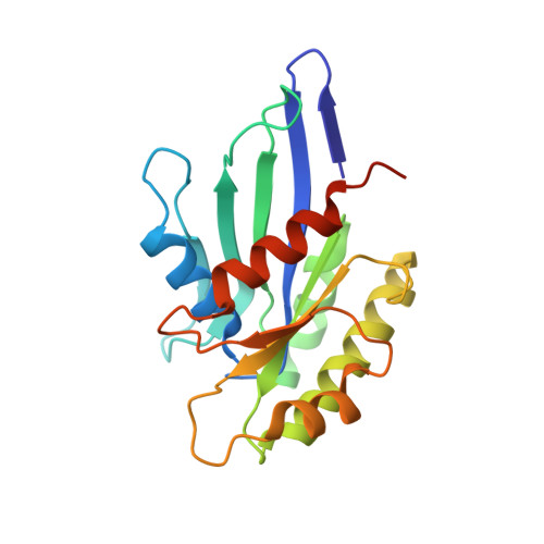
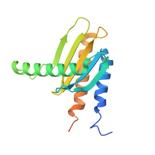
MglA functions as a three-state GTPase to control movement reversals of Myxococcus xanthus.

Galicia, C.Lhospice, S.Varela, P.F.Trapani, S.Zhang, W.Navaza, J.Herrou, J.Mignot, T.Cherfils, J.

(2019) Nat Commun 10: 5300-5300

  • DOI: https://doi.org/10.1038/s41467-019-13274-3
  • Primary Citation Related Structures: 
    6H17, 6H35, 6H5B, 6HJH, 6HJM, 6HJO

  • PubMed Abstract: 

    In Myxococcus xanthus, directed movement is controlled by pole-to-pole oscillations of the small GTPase MglA and its GAP MglB. Direction reversals require that MglA is inactivated by MglB, yet paradoxically MglA and MglB are located at opposite poles at reversal initiation. Here we report the complete MglA/MglB structural cycle combined to GAP kinetics and in vivo motility assays, which uncovers that MglA is a three-state GTPase and suggests a molecular mechanism for concerted MglA/MglB relocalizations. We show that MglA has an atypical GTP-bound state (MglA-GTP*) that is refractory to MglB and is re-sensitized by a feedback mechanism operated by MglA-GDP. By identifying and mutating the pole-binding region of MglB, we then provide evidence that the MglA-GTP* state exists in vivo. These data support a model in which MglA-GDP acts as a soluble messenger to convert polar MglA-GTP* into a diffusible MglA-GTP species that re-localizes to the opposite pole during reversals.


  • Organizational Affiliation
    • Laboratoire de Biologie et Pharmacologie Appliquée, CNRS and Ecole Normale Supérieure Paris-Saclay, Cachan, France.

Macromolecule Content 

  • Total Structure Weight: 59.24 kDa 
  • Atom Count: 3,552 
  • Modeled Residue Count: 442 
  • Deposited Residue Count: 521 
  • Unique protein chains: 2

Macromolecules

Find similar proteins by:|  3D Structure
Entity ID: 1
MoleculeChains  Sequence LengthOrganismDetailsImage
Mutual gliding-motility protein MglA201Myxococcus xanthus DK 1622Mutation(s): 0 
Gene Names: mglAMXAN_1925
UniProt
Find proteins for Q1DB04 (Myxococcus xanthus (strain DK1622))
Explore Q1DB04 
Go to UniProtKB:  Q1DB04
Entity Groups
Sequence Clusters30% Identity50% Identity70% Identity90% Identity95% Identity100% Identity
UniProt GroupQ1DB04
Sequence Annotations
Expand
Reference Sequence
Find similar proteins by:|  3D Structure
Entity ID: 2
MoleculeChains  Sequence LengthOrganismDetailsImage
MglB
B, C
160Myxococcus xanthusMutation(s): 0 
Gene Names: mglB
UniProt
Find proteins for Q50883 (Myxococcus xanthus)
Explore Q50883 
Go to UniProtKB:  Q50883
Entity Groups
Sequence Clusters30% Identity50% Identity70% Identity90% Identity95% Identity100% Identity
UniProt GroupQ50883
Sequence Annotations
Expand
Reference Sequence

Small Molecules

Ligands 5 Unique
IDChains Name / Formula / InChI Key2D Diagram3D Interactions
GSP

Query on GSP


Download:Ideal Coordinates CCD File
D [auth A]5'-GUANOSINE-DIPHOSPHATE-MONOTHIOPHOSPHATE
C10 H16 N5 O13 P3 S
XOFLBQFBSOEHOG-UUOKFMHZSA-N
EPE

Query on EPE


Download:Ideal Coordinates CCD File
F [auth A],
G [auth A]
4-(2-HYDROXYETHYL)-1-PIPERAZINE ETHANESULFONIC ACID
C8 H18 N2 O4 S
JKMHFZQWWAIEOD-UHFFFAOYSA-N
1PE

Query on 1PE


Download:Ideal Coordinates CCD File
H [auth A],
K [auth C]
PENTAETHYLENE GLYCOL
C10 H22 O6
JLFNLZLINWHATN-UHFFFAOYSA-N
PEG

Query on PEG


Download:Ideal Coordinates CCD File
I [auth B],
J [auth B],
L [auth C]
DI(HYDROXYETHYL)ETHER
C4 H10 O3
MTHSVFCYNBDYFN-UHFFFAOYSA-N
MG

Query on MG


Download:Ideal Coordinates CCD File
E [auth A]MAGNESIUM ION
Mg
JLVVSXFLKOJNIY-UHFFFAOYSA-N

Experimental Data & Validation

Experimental Data

  • Method: X-RAY DIFFRACTION
  • Resolution: 2.80 Å
  • R-Value Free:  0.255 (Depositor), 0.257 (DCC) 
  • R-Value Work:  0.199 (Depositor), 0.201 (DCC) 
  • R-Value Observed: 0.204 (Depositor) 
Space Group: P 64
Unit Cell:
Length ( Å )Angle ( ˚ )
a = 135.101α = 90
b = 135.101β = 90
c = 60.382γ = 120
Software Package:
Software NamePurpose
PHENIXrefinement
XDSdata reduction
autoPROCdata scaling
PHASERphasing

Structure Validation

View Full Validation Report


Ligand Structure Quality Assessment 


Entry History 

& Funding Information

Deposition Data


Funding OrganizationLocationGrant Number
French National Research AgencyFrance--

Revision History  (Full details and data files)

  • Version 1.0: 2019-11-27
    Type: Initial release
  • Version 1.1: 2019-12-04
    Changes: Database references
  • Version 1.2: 2020-11-11
    Changes: Refinement description, Structure summary
  • Version 1.3: 2025-04-09
    Changes: Advisory, Data collection, Database references, Refinement description, Structure summary
  • Version 1.4: 2025-10-01
    Changes: Advisory, Derived calculations