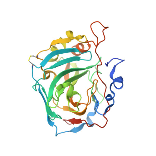
Exploring structural properties of potent human carbonic anhydrase inhibitors bearing a 4-(cycloalkylamino-1-carbonyl)benzenesulfonamide moiety.
Buemi, M.R., Di Fiore, A., De Luca, L., Angeli, A., Mancuso, F., Ferro, S., Monti, S.M., Buonanno, M., Russo, E., De Sarro, G., De Simone, G., Supuran, C.T., Gitto, R.(2018) Eur J Med Chem 163: 443-452
- PubMed: 30530195
- DOI: https://doi.org/10.1016/j.ejmech.2018.11.073
- Primary Citation of Related Structures:
6H2Z, 6H33, 6H34, 6H36, 6H37, 6H38 - PubMed Abstract:
Guided by the crystal structure of 4-(3,4-dihydroquinolin-1(2H)-ylcarbonyl)benzenesulfonamide 3 in complex with hCA II (PDB code 4Z0Q), a novel series of cycloalkylamino-1-carbonylbenzenesulfonamides was designed and synthesized. Thus, we replaced the quinoline ring with an azepine/piperidine/piperazine nucleus and introduced further modifications on cycloalkylamine nucleus by means the installation of hydrophobic/hydrophilic functionalities able to establish additional contacts in the middle area of the enzyme cavity. Among the synthesized compounds, the derivatives 7a, 7b, 8b exhibited a remarkable inhibition for hCA II and the brain-expressed hCA VII in subnanomolar range. The binding of these molecules to the target enzymes was characterized by means of a crystallographic analysis, providing a clear snapshot of the most important interactions established by this class of inhibitors into the hCA II and hCA VII catalytic site. Notably, our results showed that the benzylpiperazine tail of compound 8b is oriented both in hCA II and in hCA VII toward a poorly explored region of the active site. These features should be further investigated for the design of new isoform selective CA inhibitors.
Organizational Affiliation:
Dipartimento di Scienze Chimiche, Biologiche, Farmaceutiche ed Ambientali (CHIBIOFARAM), Università degli Studi di Messina, Viale Palatucci, Polo didattico SS, Annunziata, 98168, Messina, Italy.


