6GVU | pdb_00006gvu

NMR structure of the DNA-bound helix bundle domain from the functional pRN1 primase


Experimental Data Snapshot

  • Method: SOLUTION NMR
  • Conformers Calculated: 200 
  • Conformers Submitted: 20 
  • Selection Criteria: structures with the lowest energy 

wwPDB Validation 3D Report Full Report

Validation slider image for 6GVU

This is version 1.5 of the entry. See complete history

Literature

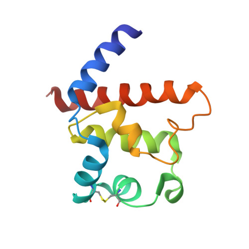
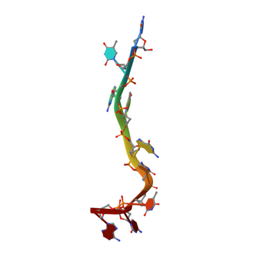
A Small Helical Bundle Prepares Primer Synthesis by Binding Two Nucleotides that Enhance Sequence-Specific Recognition of the DNA Template.

Boudet, J.Devillier, J.C.Wiegand, T.Salmon, L.Meier, B.H.Lipps, G.Allain, F.H.

(2019) Cell 176: 154-166.e13

  • DOI: https://doi.org/10.1016/j.cell.2018.11.031
  • Primary Citation Related Structures: 
    6GT7, 6GVQ, 6GVT, 6GVU

  • PubMed Abstract: 

    Primases have a fundamental role in DNA replication. They synthesize a primer that is then extended by DNA polymerases. Archaeoeukaryotic primases require for synthesis a catalytic and an accessory domain, the exact contribution of the latter being unresolved. For the pRN1 archaeal primase, this domain is a 115-amino acid helix bundle domain (HBD). Our structural investigations of this small HBD by liquid- and solid-state nuclear magnetic resonance (NMR) revealed that only the HBD binds the DNA template. DNA binding becomes sequence-specific after a major allosteric change in the HBD, triggered by the binding of two nucleotide triphosphates. The spatial proximity of the two nucleotides and the DNA template in the quaternary structure of the HBD strongly suggests that this small domain brings together the substrates to prepare the first catalytic step of primer synthesis. This efficient mechanism is likely general for all archaeoeukaryotic primases.


  • Organizational Affiliation
    • Department of Biology, Institute of Molecular Biology and Biophysics, ETH Zürich, 8093 Zürich, Switzerland. Electronic address: boudet.julien@gmail.com.

Macromolecule Content 

  • Total Structure Weight: 16.24 kDa 
  • Atom Count: 1,130 
  • Modeled Residue Count: 124 
  • Deposited Residue Count: 124 
  • Unique protein chains: 1
  • Unique nucleic acid chains: 1

Macromolecules


Find similar proteins by:|  3D Structure
Entity ID: 2
MoleculeChains  Sequence LengthOrganismDetailsImage
functional pRN1 primase115Saccharolobus islandicusMutation(s): 0 
UniProt
Find proteins for Q54324 (Saccharolobus islandicus)
Explore Q54324 
Go to UniProtKB:  Q54324
Entity Groups
Sequence Clusters30% Identity50% Identity70% Identity90% Identity95% Identity100% Identity
UniProt GroupQ54324
Sequence Annotations
Expand
Reference Sequence
Find similar nucleic acids by:  Sequence
Entity ID: 1
MoleculeChains LengthOrganismImage
DNA (5'-D(*CP*TP*GP*TP*GP*CP*TP*CP*A)-3')9Saccharolobus islandicus
Sequence Annotations
Expand
Reference Sequence

Experimental Data & Validation

Experimental Data

  • Method: SOLUTION NMR
  • Conformers Calculated: 200 
  • Conformers Submitted: 20 
  • Selection Criteria: structures with the lowest energy 

Structure Validation

View Full Validation Report


Entry History 

& Funding Information

Deposition Data


Funding OrganizationLocationGrant Number
Swiss National Science FoundationSwitzerland310030 141160

Revision History  (Full details and data files)

  • Version 1.0: 2018-12-26
    Type: Initial release
  • Version 1.1: 2019-01-09
    Changes: Data collection, Database references
  • Version 1.2: 2019-01-23
    Changes: Data collection, Database references
  • Version 1.3: 2019-05-08
    Changes: Data collection
  • Version 1.4: 2024-07-03
    Changes: Data collection, Database references, Other
  • Version 1.5: 2024-11-06
    Changes: Structure summary