6GS4 | pdb_00006gs4

Crystal structure of peptide transporter DtpA-nanobody in complex with valganciclovir


Experimental Data Snapshot

  • Method: X-RAY DIFFRACTION
  • Resolution: 2.65 Å
  • R-Value Free: 
    0.239 (Depositor), 0.241 (DCC) 
  • R-Value Work: 
    0.216 (Depositor), 0.215 (DCC) 
  • R-Value Observed: 
    0.217 (Depositor) 

Starting Model: experimental
View more details

wwPDB Validation 3D Report Full Report

Validation slider image for 6GS4

Ligand Structure Quality Assessment 


This is version 1.4 of the entry. See complete history

Literature

Structure of Prototypic Peptide Transporter DtpA from E. coli in Complex with Valganciclovir Provides Insights into Drug Binding of Human PepT1.

Ural-Blimke, Y.Flayhan, A.Strauss, J.Rantos, V.Bartels, K.Nielsen, R.Pardon, E.Steyaert, J.Kosinski, J.Quistgaard, E.M.Low, C.

(2019) J Am Chem Soc 141: 2404-2412

  • DOI: https://doi.org/10.1021/jacs.8b11343
  • Primary Citation Related Structures: 
    6GS1, 6GS4, 6GS7

  • PubMed Abstract: 

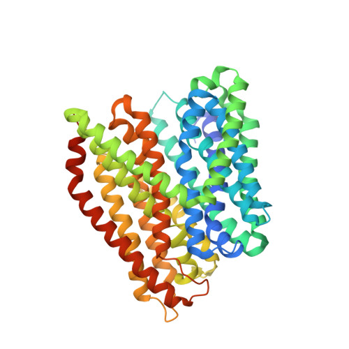
    Members of the solute carrier 15 family (SLC15) transport di- and tripeptides as well as peptidomimetic drugs across the cell membrane. Structures of bacterial homologues have provided valuable information on the binding and transport of their natural substrates, but many do not transport medically relevant drugs. In contrast, a homologue from Escherichia coli, DtpA (dipeptide and tripeptide permease), shows a high similarity to human PepT1 (SLC15A1) in terms of ligand selectivity and transports a similar set of drugs. Here, we present the crystal structure of DtpA in ligand-free form (at 3.30 Å resolution) and in complex with the antiviral prodrug valganciclovir (at 2.65 Å resolution) supported by biochemical data. We show that valganciclovir unexpectedly binds with the ganciclovir moiety mimicking the N-terminal residue of a canonical peptide substrate. On the basis of a homology model we argue that this binding mode also applies to the human PepT1 transporter. Our results provide new insights into the binding mode of prodrugs and will assist the rational design of drugs with improved absorption rates.


  • Organizational Affiliation
    • Centre for Structural Systems Biology (CSSB) , DESY and European Molecular Biology Laboratory Hamburg , Notkestrasse 85 , D-22607 Hamburg , Germany.

Macromolecule Content 

  • Total Structure Weight: 70.24 kDa 
  • Atom Count: 4,609 
  • Modeled Residue Count: 593 
  • Deposited Residue Count: 640 
  • Unique protein chains: 2

Macromolecules

Find similar proteins by:|  3D Structure
Entity ID: 1
MoleculeChains  Sequence LengthOrganismDetailsImage
Dipeptide and tripeptide permease A508Escherichia coli K-12Mutation(s): 0 
Gene Names: dtpAtppBydgRb1634JW1626
Membrane Entity: Yes 
UniProt
Find proteins for P77304 (Escherichia coli (strain K12))
Explore P77304 
Go to UniProtKB:  P77304
Entity Groups
Sequence Clusters30% Identity50% Identity70% Identity90% Identity95% Identity100% Identity
UniProt GroupP77304
Sequence Annotations
Expand
Reference Sequence
Find similar proteins by:|  3D Structure
Entity ID: 2
MoleculeChains  Sequence LengthOrganismDetailsImage
nanobodyB [auth H]132Lama glamaMutation(s): 0 

Small Molecules

Ligands 2 Unique
IDChains Name / Formula / InChI Key2D Diagram3D Interactions
LMT

Query on LMT


Download:Ideal Coordinates CCD File
D [auth A]DODECYL-BETA-D-MALTOSIDE
C24 H46 O11
NLEBIOOXCVAHBD-QKMCSOCLSA-N
F9E
(Subject of Investigation/LOI)

Query on F9E


Download:Ideal Coordinates CCD File
C [auth A][(2~{S})-2-[(2-azanyl-6-oxidanylidene-3~{H}-purin-9-yl)methoxy]-3-oxidanyl-propyl] (2~{S})-2-azanyl-3-methyl-butanoate
C14 H22 N6 O5
WPVFJKSGQUFQAP-IUCAKERBSA-N

Experimental Data & Validation

Experimental Data

  • Method: X-RAY DIFFRACTION
  • Resolution: 2.65 Å
  • R-Value Free:  0.239 (Depositor), 0.241 (DCC) 
  • R-Value Work:  0.216 (Depositor), 0.215 (DCC) 
  • R-Value Observed: 0.217 (Depositor) 
Space Group: P 21 21 21
Unit Cell:
Length ( Å )Angle ( ˚ )
a = 54.94α = 90
b = 120.19β = 90
c = 163.67γ = 90
Software Package:
Software NamePurpose
PHENIXrefinement
XDSdata reduction
XSCALEdata scaling
PHASERphasing

Structure Validation

View Full Validation Report


Ligand Structure Quality Assessment 


Entry History 

& Funding Information

Deposition Data


Funding OrganizationLocationGrant Number
European UnioniNEXT grant, project no. 653706
Swedish Research CouncilSweden621-2013-5905

Revision History  (Full details and data files)

  • Version 1.0: 2019-01-30
    Type: Initial release
  • Version 1.1: 2019-02-20
    Changes: Data collection, Database references
  • Version 1.2: 2019-11-13
    Changes: Data collection, Source and taxonomy
  • Version 1.3: 2024-01-17
    Changes: Data collection, Database references, Refinement description
  • Version 1.4: 2024-11-06
    Changes: Structure summary