6GP0 | pdb_00006gp0

Structure of mEos4b in the red fluorescent state


Experimental Data Snapshot

  • Method: X-RAY DIFFRACTION
  • Resolution: 1.50 Å
  • R-Value Free: 
    0.190 (Depositor), 0.190 (DCC) 
  • R-Value Work: 
    0.154 (Depositor), 0.154 (DCC) 
  • R-Value Observed: 
    0.155 (Depositor) 

Starting Model: experimental
View more details

wwPDB Validation 3D Report Full Report

Validation slider image for 6GP0

This is version 4.0 of the entry. See complete history

Literature

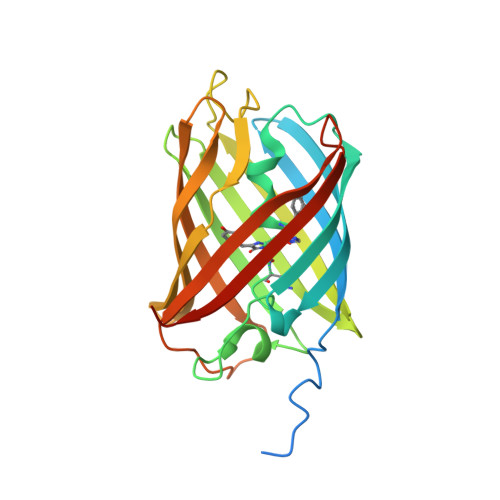
Mechanistic investigation of mEos4b reveals a strategy to reduce track interruptions in sptPALM.

De Zitter, E.Thedie, D.Monkemoller, V.Hugelier, S.Beaudouin, J.Adam, V.Byrdin, M.Van Meervelt, L.Dedecker, P.Bourgeois, D.

(2019) Nat Methods 16: 707-710

  • DOI: https://doi.org/10.1038/s41592-019-0462-3
  • Primary Citation Related Structures: 
    6GOY, 6GP0, 6GP1

  • PubMed Abstract: 

    Green-to-red photoconvertible fluorescent proteins repeatedly enter dark states, causing interrupted tracks in single-particle-tracking localization microscopy (sptPALM). We identified a long-lived dark state in photoconverted mEos4b that results from isomerization of the chromophore and efficiently absorbs cyan light. Addition of weak 488-nm light swiftly reverts this dark state to the fluorescent state. This strategy largely eliminates slow blinking and enables the recording of longer tracks in sptPALM with minimum effort.


  • Organizational Affiliation
    • Department of Chemistry, KU Leuven, Heverlee, Belgium.

Macromolecule Content 

  • Total Structure Weight: 29.55 kDa 
  • Atom Count: 2,304 
  • Modeled Residue Count: 224 
  • Deposited Residue Count: 259 
  • Unique protein chains: 1

Macromolecules

Find similar proteins by:|  3D Structure
Entity ID: 1
MoleculeChains  Sequence LengthOrganismDetailsImage
Green to red photoconvertible GFP-like protein EosFP257Lobophyllia hemprichiiMutation(s): 1 
UniProt
Find proteins for Q5S6Z9 (Lobophyllia hemprichii)
Explore Q5S6Z9 
Go to UniProtKB:  Q5S6Z9
Entity Groups
Sequence Clusters30% Identity50% Identity70% Identity90% Identity95% Identity100% Identity
UniProt GroupQ5S6Z9
Sequence Annotations
Expand
Reference Sequence

Small Molecules

Modified Residues  2 Unique
IDChains TypeFormula2D DiagramParent
5SQ
Query on 5SQ
A
L-PEPTIDE LINKINGC17 H17 N5 O4HIS, TYR, GLY
NFA
Query on NFA
A
L-PEPTIDE LINKINGC9 H12 N2 OPHE

Experimental Data & Validation

Experimental Data

  • Method: X-RAY DIFFRACTION
  • Resolution: 1.50 Å
  • R-Value Free:  0.190 (Depositor), 0.190 (DCC) 
  • R-Value Work:  0.154 (Depositor), 0.154 (DCC) 
  • R-Value Observed: 0.155 (Depositor) 
Space Group: P 21 21 21
Unit Cell:
Length ( Å )Angle ( ˚ )
a = 38.61α = 90
b = 57.9β = 90
c = 102.64γ = 90
Software Package:
Software NamePurpose
XDSdata reduction
XSCALEdata scaling
PHENIXrefinement
PDB_EXTRACTdata extraction
PHENIXphasing

Structure Validation

View Full Validation Report


Entry History 

& Funding Information

Deposition Data


Funding OrganizationLocationGrant Number
Research Foundation - FlandersBelgium--
Grenoble Instruct-ERIC CenterFranceUMS 3518 CNRS-CEA-UGA-EMBL
FranceANR-10-INBS-05-02
FranceANR-10-LABX-49-01
France--

Revision History  (Full details and data files)

  • Version 1.0: 2019-05-22
    Type: Initial release
  • Version 1.1: 2019-07-17
    Changes: Data collection, Database references
  • Version 1.2: 2019-08-07
    Changes: Data collection, Database references
  • Version 2.0: 2021-10-13
    Changes: Advisory, Atomic model, Author supporting evidence, Data collection, Database references, Derived calculations, Non-polymer description, Polymer sequence, Source and taxonomy, Structure summary
  • Version 3.0: 2023-11-15
    Changes: Atomic model, Data collection, Derived calculations
  • Version 3.1: 2024-01-17
    Changes: Refinement description
  • Version 4.0: 2025-07-16
    Changes: Advisory, Atomic model, Database references, Derived calculations, Structure summary