6GO1 | pdb_00006go1

Crystal Structure of a Bacillus anthracis peptidoglycan deacetylase


Experimental Data Snapshot

  • Method: X-RAY DIFFRACTION
  • Resolution: 2.59 Å
  • R-Value Free: 
    0.245 (Depositor), 0.246 (DCC) 
  • R-Value Work: 
    0.183 (Depositor), 0.183 (DCC) 
  • R-Value Observed: 
    0.186 (Depositor) 

Starting Model: experimental
View more details

wwPDB Validation 3D Report Full Report

Validation slider image for 6GO1

This is version 1.2 of the entry. See complete history

Literature

The putative polysaccharide deacetylase Ba0331: cloning, expression, crystallization and structure determination.

Andreou, A.Giastas, P.Arnaouteli, S.Tzanodaskalaki, M.Tzartos, S.J.Bethanis, K.Bouriotis, V.Eliopoulos, E.E.

(2019) Acta Crystallogr F Struct Biol Commun 75: 312-320

  • DOI: https://doi.org/10.1107/S2053230X19001766
  • Primary Citation Related Structures: 
    6GO1

  • PubMed Abstract: 

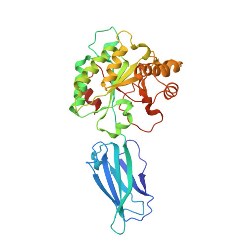
    Ba0331 is a putative polysaccharide deacetylase from Bacillus anthracis, the etiological agent of the disease anthrax, that contributes to adaptation of the bacterium under extreme conditions and to maintenance of the cell shape. In the present study, the crystal structure of Ba0331 was determined at 2.6 Å resolution. The structure consists of two domains: a fibronectin type 3-like (Fn3-like) domain and a NodB catalytic domain. The latter is present in all carbohydrate esterase family 4 enzymes, while a comparative analysis of the Fn3-like domain revealed structural plasticity despite the retention of the conserved Fn3-like domain characteristics.


  • Organizational Affiliation
    • Department of Biotechnology, Agricultural University of Athens, Iera Odos 75, 11855 Athens, Greece.

Macromolecule Content 

  • Total Structure Weight: 74.4 kDa 
  • Atom Count: 5,294 
  • Modeled Residue Count: 636 
  • Deposited Residue Count: 636 
  • Unique protein chains: 1

Macromolecules

Find similar proteins by:|  3D Structure
Entity ID: 1
MoleculeChains  Sequence LengthOrganismDetailsImage
Polysaccharide deacetylase-like protein
A, B
318Bacillus anthracisMutation(s): 0 
Gene Names: yxkHBA_0331BASH2_05488
UniProt
Find proteins for A0A3P1TYX8 (Bacillus anthracis)
Explore A0A3P1TYX8 
Go to UniProtKB:  A0A3P1TYX8
Entity Groups
Sequence Clusters30% Identity50% Identity70% Identity90% Identity95% Identity100% Identity
UniProt GroupA0A3P1TYX8
Sequence Annotations
Expand
Reference Sequence

Small Molecules

Ligands 4 Unique
IDChains Name / Formula / InChI Key2D Diagram3D Interactions
SO4

Query on SO4


Download:Ideal Coordinates CCD File
G [auth A]SULFATE ION
O4 S
QAOWNCQODCNURD-UHFFFAOYSA-L
ZN

Query on ZN


Download:Ideal Coordinates CCD File
C [auth A],
H [auth B]
ZINC ION
Zn
PTFCDOFLOPIGGS-UHFFFAOYSA-N
EDO

Query on EDO


Download:Ideal Coordinates CCD File
E [auth A],
F [auth A],
J [auth B],
K [auth B]
1,2-ETHANEDIOL
C2 H6 O2
LYCAIKOWRPUZTN-UHFFFAOYSA-N
ACT

Query on ACT


Download:Ideal Coordinates CCD File
D [auth A],
I [auth B]
ACETATE ION
C2 H3 O2
QTBSBXVTEAMEQO-UHFFFAOYSA-M

Experimental Data & Validation

Experimental Data

  • Method: X-RAY DIFFRACTION
  • Resolution: 2.59 Å
  • R-Value Free:  0.245 (Depositor), 0.246 (DCC) 
  • R-Value Work:  0.183 (Depositor), 0.183 (DCC) 
  • R-Value Observed: 0.186 (Depositor) 
Space Group: P 1 21 1
Unit Cell:
Length ( Å )Angle ( ˚ )
a = 41.723α = 90
b = 211.795β = 91.82
c = 46.784γ = 90
Software Package:
Software NamePurpose
XDSdata reduction
XDSdata scaling
PHASERphasing
PHENIXrefinement
PDB_EXTRACTdata extraction

Structure Validation

View Full Validation Report


Entry History 

Deposition Data

Revision History  (Full details and data files)

  • Version 1.0: 2019-04-10
    Type: Initial release
  • Version 1.1: 2019-04-17
    Changes: Data collection, Database references
  • Version 1.2: 2024-01-17
    Changes: Data collection, Database references, Derived calculations, Refinement description