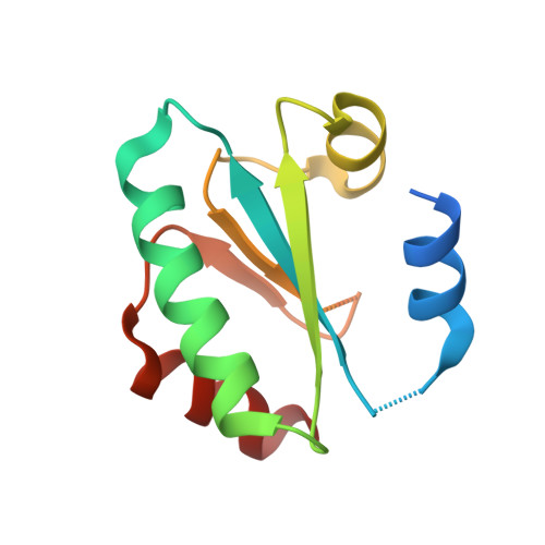
6GND

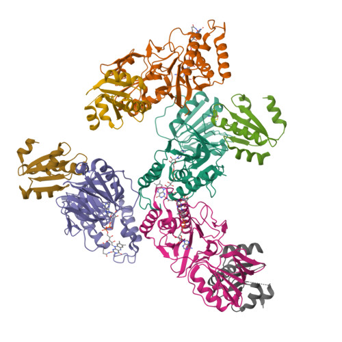
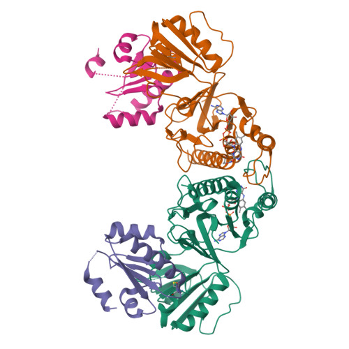
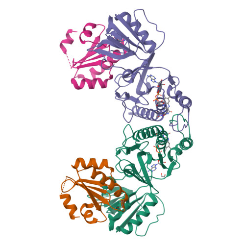
Crystal structure of the complex of a Ferredoxin-Flavin Thioredoxin Reductase and a Thioredoxin from Clostridium acetobutylicum at 2.9 A resolution


Experimental Data Snapshot

  • Method: X-RAY DIFFRACTION
  • Resolution: 2.89 Å
  • R-Value Free: 0.279 
  • R-Value Work: 0.235 
  • R-Value Observed: 0.237 

Starting Model: experimental
View more details

wwPDB Validation   3D Report Full Report


Ligand Structure Quality Assessment 


This is version 1.2 of the entry. See complete history


Literature

Ferredoxin-linked flavoenzyme defines a family of pyridine nucleotide-independent thioredoxin reductases.

Buey, R.M.Fernandez-Justel, D.de Pereda, J.M.Revuelta, J.L.Schurmann, P.Buchanan, B.B.Balsera, M.

(2018) Proc Natl Acad Sci U S A 115: 12967-12972

  • DOI: https://doi.org/10.1073/pnas.1812781115
  • Primary Citation of Related Structures:  
    6GN9, 6GNA, 6GNB, 6GNC, 6GND

  • PubMed Abstract: 

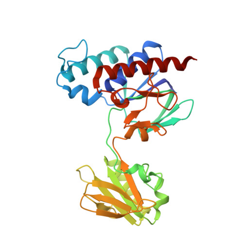
    Ferredoxin-dependent thioredoxin reductase was identified 35 y ago in the fermentative bacterium Clostridium pasteurianum [Hammel KE, Cornwell KL, Buchanan BB (1983) Proc Natl Acad Sci USA 80:3681-3685]. The enzyme, a flavoprotein, was strictly dependent on ferredoxin as reductant and was inactive with either NADPH or NADH. This early work has not been further pursued. We have recently reinvestigated the problem and confirmed that the enzyme, here designated ferredoxin-dependent flavin thioredoxin reductase (FFTR), is a flavoprotein. The enzyme differs from ferredoxin-thioredoxin reductase (FTR), which has a signature [4Fe-4S] cluster, but shows structural similarities to NADP-dependent thioredoxin reductase (NTR). Comparative amino acid sequence analysis showed that FFTR is present in a number of clostridial species, some of which lack both FTR and an archetypal NTR. We have isolated, crystallized, and determined the structural properties of FFTR from a member of this group, Clostridium acetobutylicum , both alone and in complex with Trx. The structures showed an elongated FFTR homodimer, each monomer comprising two Rossmann domains and a noncovalently bound FAD cofactor that exposes the isoalloxazine ring to the solvent. The FFTR structures revealed an alternative domain organization compared with NTR that enables the enzyme to accommodate Fdx rather than NADPH. The results suggest that FFTR exists in a range of conformations with varying degrees of domain separation in solution and that the stacking between the two redox-active groups for the transfer of reducing equivalents results in a profound structural reorganization. A mechanism in accord with the findings is proposed.


  • Organizational Affiliation

    Metabolic Engineering Group, Departamento de Microbiología y Genética, Universidad de Salamanca, 37007 Salamanca, Spain.


Macromolecules
Find similar proteins by:  (by identity cutoff)  |  3D Structure
Entity ID: 1
MoleculeChains Sequence LengthOrganismDetailsImage
Thioredoxin reductase
A, C, E, G
288Clostridium acetobutylicum ATCC 824Mutation(s): 1 
Gene Names: CA_C3082
UniProt
Find proteins for Q97EM8 (Clostridium acetobutylicum (strain ATCC 824 / DSM 792 / JCM 1419 / IAM 19013 / LMG 5710 / NBRC 13948 / NRRL B-527 / VKM B-1787 / 2291 / W))
Explore Q97EM8 
Go to UniProtKB:  Q97EM8
Entity Groups  
Sequence Clusters30% Identity50% Identity70% Identity90% Identity95% Identity100% Identity
UniProt GroupQ97EM8
Sequence Annotations
Expand
  • Reference Sequence
Find similar proteins by:  (by identity cutoff)  |  3D Structure
Entity ID: 2
MoleculeChains Sequence LengthOrganismDetailsImage
Thioredoxin
B, D, F, H
108Clostridium acetobutylicum ATCC 824Mutation(s): 1 
Gene Names: CA_C3083
UniProt
Find proteins for Q97EM7 (Clostridium acetobutylicum (strain ATCC 824 / DSM 792 / JCM 1419 / IAM 19013 / LMG 5710 / NBRC 13948 / NRRL B-527 / VKM B-1787 / 2291 / W))
Explore Q97EM7 
Go to UniProtKB:  Q97EM7
Entity Groups  
Sequence Clusters30% Identity50% Identity70% Identity90% Identity95% Identity100% Identity
UniProt GroupQ97EM7
Sequence Annotations
Expand
  • Reference Sequence
Experimental Data & Validation

Experimental Data

  • Method: X-RAY DIFFRACTION
  • Resolution: 2.89 Å
  • R-Value Free: 0.279 
  • R-Value Work: 0.235 
  • R-Value Observed: 0.237 
  • Space Group: C 1 2 1
Unit Cell:
Length ( Å )Angle ( ˚ )
a = 115.23α = 90
b = 174.35β = 119.8
c = 114.304γ = 90
Software Package:
Software NamePurpose
PHENIXrefinement
XDSdata reduction
XSCALEdata scaling
PHASERphasing

Structure Validation

View Full Validation Report


Ligand Structure Quality Assessment 


Entry History & Funding Information

Deposition Data


Funding OrganizationLocationGrant Number
Ministry of Economy and CompetitivenessSpainBFU2016-80343-P

Revision History  (Full details and data files)

  • Version 1.0: 2018-12-12
    Type: Initial release
  • Version 1.1: 2019-01-02
    Changes: Data collection, Database references
  • Version 1.2: 2024-01-17
    Changes: Data collection, Database references, Refinement description