6GKY

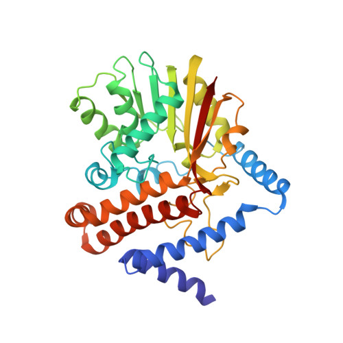
Crystal structure of Coclaurine N-Methyltransferase (CNMT) bound to N-methylheliamine and SAH


Experimental Data Snapshot

  • Method: X-RAY DIFFRACTION
  • Resolution: 2.85 Å
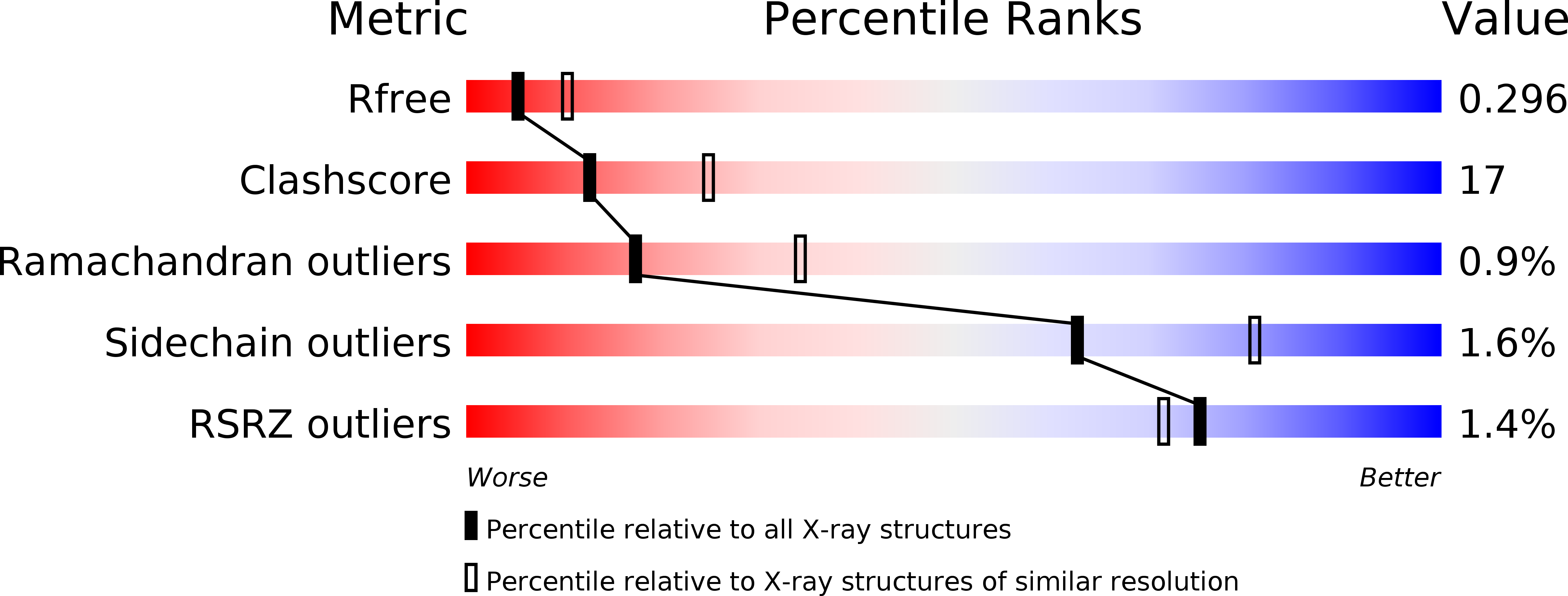
  • R-Value Free: 0.295 
  • R-Value Work: 0.215 
  • R-Value Observed: 0.219 

wwPDB Validation   3D Report Full Report


Ligand Structure Quality Assessment 


This is version 1.3 of the entry. See complete history


Literature

Structure and Biocatalytic Scope of Coclaurine N-Methyltransferase.

Bennett, M.R.Thompson, M.L.Shepherd, S.A.Dunstan, M.S.Herbert, A.J.Smith, D.R.M.Cronin, V.A.Menon, B.R.K.Levy, C.Micklefield, J.

(2018) Angew Chem Int Ed Engl 57: 10600-10604

  • DOI: https://doi.org/10.1002/anie.201805060
  • Primary Citation of Related Structures:  
    6GKV, 6GKY, 6GKZ

  • PubMed Abstract: 

    Benzylisoquinoline alkaloids (BIAs) are a structurally diverse family of plant secondary metabolites, which have been exploited to develop analgesics, antibiotics, antitumor agents, and other therapeutic agents. Biosynthesis of BIAs proceeds via a common pathway from tyrosine to (S)-reticulene at which point the pathway diverges. Coclaurine N-methyltransferase (CNMT) is a key enzyme in the pathway to (S)-reticulene, installing the N-methyl substituent that is essential for the bioactivity of many BIAs. In this paper, we describe the first crystal structure of CNMT which, along with mutagenesis studies, defines the enzymes active site architecture. The specificity of CNMT was also explored with a range of natural and synthetic substrates as well as co-factor analogues. Knowledge from this study could be used to generate improved CNMT variants required to produce BIAs or synthetic derivatives.


  • Organizational Affiliation

    School of Chemistry, Manchester Institute of Biotechnology, The University of Manchester, 131 Princess Street, Manchester, M1 7DN, UK.


Macromolecules
Find similar proteins by:  (by identity cutoff)  |  3D Structure
Entity ID: 1
MoleculeChains Sequence LengthOrganismDetailsImage
Coclaurine N-methyltransferase
A, B
351Coptis japonicaMutation(s): 0 
Gene Names: cnmt
EC: 2.1.1.115
UniProt
Find proteins for Q948P7 (Coptis japonica)
Explore Q948P7 
Go to UniProtKB:  Q948P7
Entity Groups  
Sequence Clusters30% Identity50% Identity70% Identity90% Identity95% Identity100% Identity
UniProt GroupQ948P7
Sequence Annotations
Expand
  • Reference Sequence
Experimental Data & Validation

Experimental Data

  • Method: X-RAY DIFFRACTION
  • Resolution: 2.85 Å
  • R-Value Free: 0.295 
  • R-Value Work: 0.215 
  • R-Value Observed: 0.219 
  • Space Group: P 21 21 21
Unit Cell:
Length ( Å )Angle ( ˚ )
a = 48.295α = 90
b = 95.957β = 90
c = 154.567γ = 90
Software Package:
Software NamePurpose
PHENIXrefinement
PDB_EXTRACTdata extraction
XDSdata reduction
X-Areadata scaling
PHENIXphasing

Structure Validation

View Full Validation Report


Ligand Structure Quality Assessment 


Entry History & Funding Information

Deposition Data


Funding OrganizationLocationGrant Number
United Kingdom--

Revision History  (Full details and data files)

  • Version 1.0: 2018-06-06
    Type: Initial release
  • Version 1.1: 2018-06-13
    Changes: Data collection, Derived calculations
  • Version 1.2: 2018-08-29
    Changes: Data collection, Database references
  • Version 1.3: 2024-05-15
    Changes: Data collection, Database references