6GJY

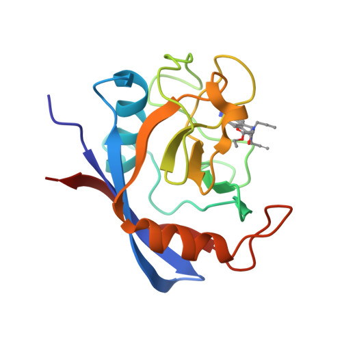
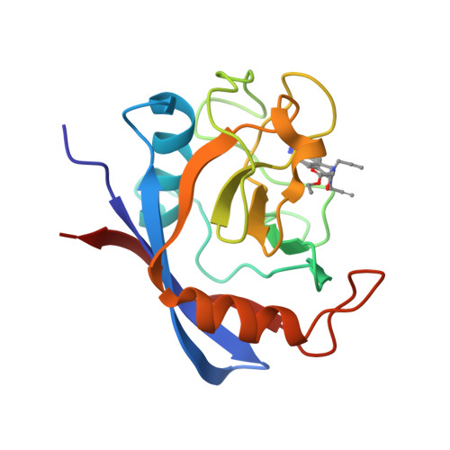
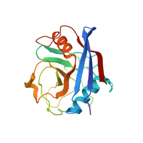
Cyclophilin A complexed with tri-vector ligand 5.


Experimental Data Snapshot

  • Method: X-RAY DIFFRACTION
  • Resolution: 1.29 Å
  • R-Value Free: 0.188 
  • R-Value Work: 0.164 
  • R-Value Observed: 0.165 

Starting Model: experimental
View more details

wwPDB Validation   3D Report Full Report


Ligand Structure Quality Assessment 


This is version 1.2 of the entry. See complete history


Literature

A computationally designed binding mode flip leads to a novel class of potent tri-vector cyclophilin inhibitors.

De Simone, A.Georgiou, C.Ioannidis, H.Gupta, A.A.Juarez-Jimenez, J.Doughty-Shenton, D.Blackburn, E.A.Wear, M.A.Richards, J.P.Barlow, P.N.Carragher, N.Walkinshaw, M.D.Hulme, A.N.Michel, J.

(2019) Chem Sci 10: 542-547

  • DOI: https://doi.org/10.1039/c8sc03831g
  • Primary Citation of Related Structures:  
    6GJI, 6GJJ, 6GJL, 6GJM, 6GJN, 6GJP, 6GJR, 6GJY

  • PubMed Abstract: 

    Cyclophilins (Cyps) are a major family of drug targets that are challenging to prosecute with small molecules because the shallow nature and high degree of conservation of the active site across human isoforms offers limited opportunities for potent and selective inhibition. Herein a computational approach based on molecular dynamics simulations and free energy calculations was combined with biophysical assays and X-ray crystallography to explore a flip in the binding mode of a reported urea-based Cyp inhibitor. This approach enabled access to a distal pocket that is poorly conserved among key Cyp isoforms, and led to the discovery of a new family of sub-micromolar cell-active inhibitors that offer unprecedented opportunities for the development of next-generation drug therapies based on Cyp inhibition. The computational approach is applicable to a broad range of organic functional groups and could prove widely enabling in molecular design.


  • Organizational Affiliation

    University of Edinburgh , Joseph Black Building, King's Buildings, David Brewster Road , Edinburgh , Scotland EH9 3FJ , UK . Email: mail@julienmichel.net.


Macromolecules
Find similar proteins by:  (by identity cutoff)  |  3D Structure
Entity ID: 1
MoleculeChains Sequence LengthOrganismDetailsImage
Peptidyl-prolyl cis-trans isomerase A165Homo sapiensMutation(s): 0 
Gene Names: PPIACYPA
EC: 5.2.1.8
UniProt & NIH Common Fund Data Resources
Find proteins for P62937 (Homo sapiens)
Explore P62937 
Go to UniProtKB:  P62937
PHAROS:  P62937
GTEx:  ENSG00000196262 
Entity Groups  
Sequence Clusters30% Identity50% Identity70% Identity90% Identity95% Identity100% Identity
UniProt GroupP62937
Sequence Annotations
Expand
  • Reference Sequence
Small Molecules
Ligands 1 Unique
IDChains Name / Formula / InChI Key2D Diagram3D Interactions
F1Z
Query on F1Z

Download Ideal Coordinates CCD File 
B [auth A]ethyl 2-[[(4-aminophenyl)methyl-prop-2-ynyl-carbamoyl]amino]ethanoate
C15 H19 N3 O3
XRVDTFOBXOWARD-UHFFFAOYSA-N
Experimental Data & Validation

Experimental Data

  • Method: X-RAY DIFFRACTION
  • Resolution: 1.29 Å
  • R-Value Free: 0.188 
  • R-Value Work: 0.164 
  • R-Value Observed: 0.165 
  • Space Group: P 21 21 21
Unit Cell:
Length ( Å )Angle ( ˚ )
a = 42.59α = 90
b = 52.84β = 90
c = 87.98γ = 90
Software Package:
Software NamePurpose
REFMACrefinement
xia2data reduction
Aimlessdata scaling
REFMACphasing

Structure Validation

View Full Validation Report


Ligand Structure Quality Assessment 


Entry History & Funding Information

Deposition Data


Funding OrganizationLocationGrant Number
European Research CouncilUnited KingdomNo. 336289

Revision History  (Full details and data files)

  • Version 1.0: 2018-11-07
    Type: Initial release
  • Version 1.1: 2019-02-20
    Changes: Data collection, Database references
  • Version 1.2: 2024-01-17
    Changes: Data collection, Database references, Refinement description