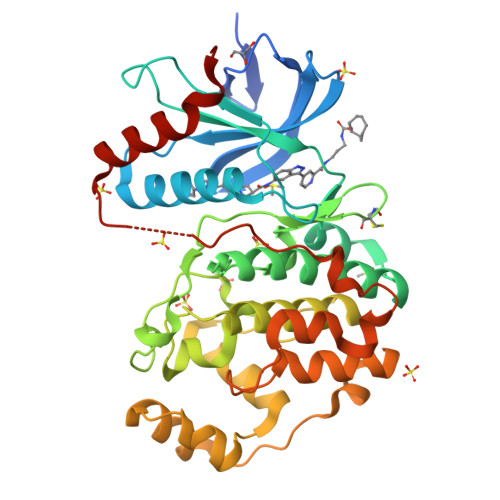
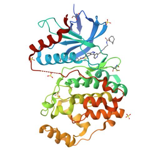
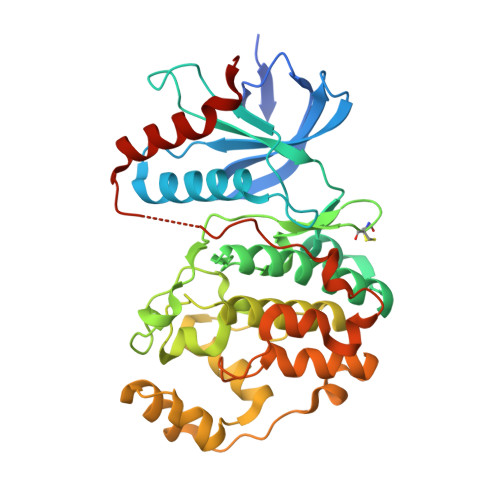
6GJD | pdb_00006gjd

Erk2 signalling protein


Experimental Data Snapshot

  • Method: X-RAY DIFFRACTION
  • Resolution: 1.58 Å
  • R-Value Free: 
    0.210 (Depositor), 0.210 (DCC) 
  • R-Value Work: 
    0.167 (Depositor), 0.180 (DCC) 
  • R-Value Observed: 
    0.169 (Depositor) 

wwPDB Validation   3D Report Full Report


Ligand Structure Quality Assessment 

Created with Raphaël 2.3.0Worse 01 BetterLigand structure goodness of fit to experimental dataBest fitted F0EClick on this verticalbar to view details

This is version 1.1 of the entry. See complete history


Literature

Quantitation of ERK1/2 inhibitor cellular target occupancies with a reversible slow off-rate probe.

Lebraud, H.Surova, O.Courtin, A.O'Reilly, M.Valenzano, C.R.Nordlund, P.Heightman, T.D.

(2018) Chem Sci 9: 8608-8618

  • DOI: https://doi.org/10.1039/c8sc02754d
  • Primary Citation of Related Structures:  
    6GJB, 6GJD

  • PubMed Abstract: 

    Target engagement is a key concept in drug discovery and its direct measurement can provide a quantitative understanding of drug efficacy and/or toxicity. Failure to demonstrate target occupancy in relevant cells and tissues has been recognised as a contributing factor to the low success rate of clinical drug development. Several techniques are emerging to quantify target engagement in cells; however, in situ measurements remain challenging, mainly due to technical limitations. Here, we report the development of a non-covalent clickable probe, based on SCH772984, a slow off-rate ERK1/2 inhibitor, which enabled efficient pull down of ERK1/2 protein via click reaction with tetrazine tagged agarose beads. This was used in a competition setting to measure relative target occupancy by selected ERK1/2 inhibitors. As a reference we used the cellular thermal shift assay, a label-free biophysical assay relying solely on ligand-induced thermodynamic stabilization of proteins. To validate the EC 50 values measured by both methods, the results were compared with IC 50 data for the phosphorylation of RSK, a downstream substrate of ERK1/2 used as a functional biomarker of ERK1/2 inhibition. We showed that a slow off-rate reversible probe can be used to efficiently pull down cellular proteins, significantly extending the potential of the approach beyond the need for covalent or photoaffinity warheads.


  • Organizational Affiliation

    Astex Pharmaceuticals , 436 Cambridge Science Park , Cambridge , CB4 0QA , UK . Email: honorine.lebraud@astx.com ; Email: tom.heightman@astx.com.


Macromolecules
Find similar proteins by:  (by identity cutoff)  |  3D Structure
Entity ID: 1
MoleculeChains Sequence LengthOrganismDetailsImage
Mitogen-activated protein kinase 1368Homo sapiensMutation(s): 0 
Gene Names: MAPK1ERK2PRKM1PRKM2
EC: 2.7.11.24
UniProt & NIH Common Fund Data Resources
Find proteins for P28482 (Homo sapiens)
Explore P28482 
Go to UniProtKB:  P28482
PHAROS:  P28482
GTEx:  ENSG00000100030 
Entity Groups  
Sequence Clusters30% Identity50% Identity70% Identity90% Identity95% Identity100% Identity
UniProt GroupP28482
Sequence Annotations
Expand
  • Reference Sequence
Small Molecules
Ligands 4 Unique
IDChains Name / Formula / InChI Key2D Diagram3D Interactions
F0E (Subject of Investigation/LOI)
Query on F0E

Download Ideal Coordinates CCD File 
L [auth A]cyclooctyl ~{N}-[3-[[4-[5-[[(3~{R})-1-[2-oxidanylidene-2-[4-(4-pyrimidin-2-ylphenyl)piperazin-1-yl]ethyl]pyrrolidin-3-yl]carbonylamino]-1~{H}-indazol-3-yl]pyridin-2-yl]carbonylamino]propyl]carbamate
C46 H55 N11 O5
FWLOTDDRDKLCPO-UUWRZZSWSA-N
SO4
Query on SO4

Download Ideal Coordinates CCD File 
B [auth A]
F [auth A]
G [auth A]
H [auth A]
I [auth A]
SULFATE ION
O4 S
QAOWNCQODCNURD-UHFFFAOYSA-L
GOL
Query on GOL

Download Ideal Coordinates CCD File 
E [auth A]GLYCEROL
C3 H8 O3
PEDCQBHIVMGVHV-UHFFFAOYSA-N
DMS
Query on DMS

Download Ideal Coordinates CCD File 
C [auth A],
D [auth A]
DIMETHYL SULFOXIDE
C2 H6 O S
IAZDPXIOMUYVGZ-UHFFFAOYSA-N
Modified Residues  1 Unique
IDChains TypeFormula2D DiagramParent
CME
Query on CME
A
L-PEPTIDE LINKINGC5 H11 N O3 S2CYS
Experimental Data & Validation

Experimental Data

  • Method: X-RAY DIFFRACTION
  • Resolution: 1.58 Å
  • R-Value Free:  0.210 (Depositor), 0.210 (DCC) 
  • R-Value Work:  0.167 (Depositor), 0.180 (DCC) 
  • R-Value Observed: 0.169 (Depositor) 
Space Group: P 1 21 1
Unit Cell:
Length ( Å )Angle ( ˚ )
a = 48.77α = 90
b = 70.996β = 109.42
c = 60.12γ = 90
Software Package:
Software NamePurpose
BUSTERrefinement
XDSdata reduction
Aimlessdata scaling
BUSTERphasing

Structure Validation

View Full Validation Report


Ligand Structure Quality Assessment 

Created with Raphaël 2.3.0Worse 01 BetterLigand structure goodness of fit to experimental dataBest fitted F0EClick on this verticalbar to view details

Entry History 

Deposition Data

  • Released Date: 2019-01-02 
  • Deposition Author(s): O'Reilly, M.

Revision History  (Full details and data files)

  • Version 1.0: 2019-01-02
    Type: Initial release
  • Version 1.1: 2024-10-23
    Changes: Data collection, Database references, Structure summary