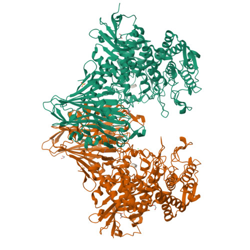
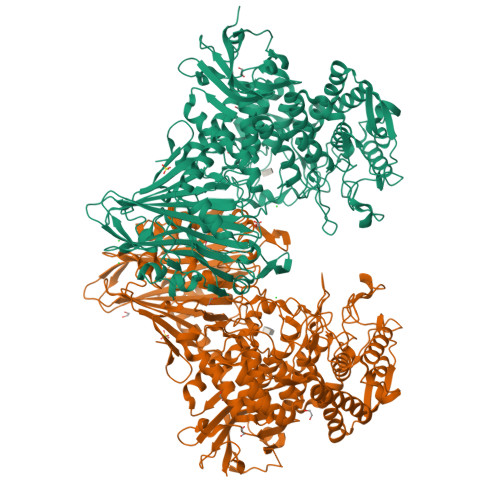
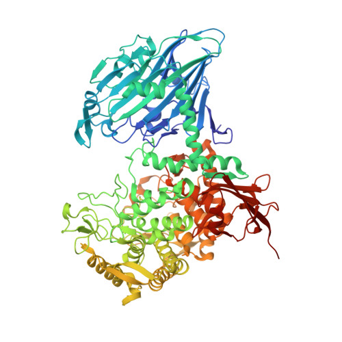
6GH3

Paenibacillus sp. YM1 laminaribiose phosphorylase with alpha-man-1-phosphate bound


Experimental Data Snapshot

  • Method: X-RAY DIFFRACTION
  • Resolution: 1.82 Å
  • R-Value Free: 0.218 
  • R-Value Work: 0.194 
  • R-Value Observed: 0.195 

Starting Model: experimental
View more details

wwPDB Validation   3D Report Full Report


Ligand Structure Quality Assessment 


This is version 1.3 of the entry. See complete history


Literature

Unravelling the Specificity of Laminaribiose Phosphorylase from Paenibacillus sp. YM-1 towards Donor Substrates Glucose/Mannose 1-Phosphate by Using X-ray Crystallography and Saturation Transfer Difference NMR Spectroscopy.

Kuhaudomlarp, S.Walpole, S.Stevenson, C.E.M.Nepogodiev, S.A.Lawson, D.M.Angulo, J.Field, R.A.

(2019) Chembiochem 20: 181-192

  • DOI: https://doi.org/10.1002/cbic.201800260
  • Primary Citation of Related Structures:  
    6GGY, 6GH2, 6GH3

  • PubMed Abstract: 

    Glycoside phosphorylases (GPs) carry out a reversible phosphorolysis of carbohydrates into oligosaccharide acceptors and the corresponding sugar 1-phosphates. The reversibility of the reaction enables the use of GPs as biocatalysts for carbohydrate synthesis. Glycosyl hydrolase family 94 (GH94), which only comprises GPs, is one of the most studied GP families that have been used as biocatalysts for carbohydrate synthesis, in academic research and in industrial production. Understanding the mechanism of GH94 enzymes is a crucial step towards enzyme engineering to improve and expand the applications of these enzymes in synthesis. In this work with a GH94 laminaribiose phosphorylase from Paenibacillus sp. YM-1 (PsLBP), we have demonstrated an enzymatic synthesis of disaccharide 1 (β-d-mannopyranosyl-(1→3)-d-glucopyranose) by using a natural acceptor glucose and noncognate donor substrate α-mannose 1-phosphate (Man1P). To investigate how the enzyme recognises different sugar 1-phosphates, the X-ray crystal structures of PsLBP in complex with Glc1P and Man1P have been solved, providing the first molecular detail of the recognition of a noncognate donor substrate by GPs, which revealed the importance of hydrogen bonding between the active site residues and hydroxy groups at C2, C4, and C6 of sugar 1-phosphates. Furthermore, we used saturation transfer difference NMR spectroscopy to support crystallographic studies on the sugar 1-phosphates, as well as to provide further insights into the PsLBP recognition of the acceptors and disaccharide products.


  • Organizational Affiliation

    Department of Biological Chemistry, John Innes Centre, Norwich Research Park, Norwich, NR4 7UH, UK.


Macromolecules
Find similar proteins by:  (by identity cutoff)  |  3D Structure
Entity ID: 1
MoleculeChains Sequence LengthOrganismDetailsImage
Laminaribiose phosphorylase
A, B
913Paenibacillus sp. YM1Mutation(s): 0 
Gene Names: lbpA
EC: 2.4.1.31
UniProt
Find proteins for D7UT17 (Paenibacillus sp. YM1)
Explore D7UT17 
Go to UniProtKB:  D7UT17
Entity Groups  
Sequence Clusters30% Identity50% Identity70% Identity90% Identity95% Identity100% Identity
UniProt GroupD7UT17
Sequence Annotations
Expand
  • Reference Sequence
Small Molecules
Ligands 4 Unique
IDChains Name / Formula / InChI Key2D Diagram3D Interactions
M1P
Query on M1P

Download Ideal Coordinates CCD File 
D [auth A],
M [auth B]
1-O-phosphono-alpha-D-mannopyranose
C6 H13 O9 P
HXXFSFRBOHSIMQ-RWOPYEJCSA-N
SO4
Query on SO4

Download Ideal Coordinates CCD File 
C [auth A],
L [auth B]
SULFATE ION
O4 S
QAOWNCQODCNURD-UHFFFAOYSA-L
EDO
Query on EDO

Download Ideal Coordinates CCD File 
E [auth A]
F [auth A]
G [auth A]
H [auth A]
I [auth A]
E [auth A],
F [auth A],
G [auth A],
H [auth A],
I [auth A],
N [auth B],
O [auth B],
P [auth B],
Q [auth B],
R [auth B],
S [auth B]
1,2-ETHANEDIOL
C2 H6 O2
LYCAIKOWRPUZTN-UHFFFAOYSA-N
CL
Query on CL

Download Ideal Coordinates CCD File 
J [auth A],
K [auth A],
T [auth B],
U [auth B]
CHLORIDE ION
Cl
VEXZGXHMUGYJMC-UHFFFAOYSA-M
Experimental Data & Validation

Experimental Data

  • Method: X-RAY DIFFRACTION
  • Resolution: 1.82 Å
  • R-Value Free: 0.218 
  • R-Value Work: 0.194 
  • R-Value Observed: 0.195 
  • Space Group: P 41 21 2
Unit Cell:
Length ( Å )Angle ( ˚ )
a = 146.57α = 90
b = 146.57β = 90
c = 222.227γ = 90
Software Package:
Software NamePurpose
XDSdata reduction
Aimlessdata scaling
REFMACrefinement
PDB_EXTRACTdata extraction
REFMACphasing

Structure Validation

View Full Validation Report


Ligand Structure Quality Assessment 


Entry History & Funding Information

Deposition Data


Funding OrganizationLocationGrant Number
Biotechnology and Biological Sciences Research CouncilUnited KingdomBB/J004561/1
Biotechnology and Biological Sciences Research CouncilUnited KingdomBB/P010660/1
Engineering and Physical Sciences Research CouncilUnited KingdomBB/M02903411

Revision History  (Full details and data files)

  • Version 1.0: 2018-06-13
    Type: Initial release
  • Version 1.1: 2019-04-24
    Changes: Data collection, Database references
  • Version 1.2: 2020-07-29
    Type: Remediation
    Reason: Carbohydrate remediation
    Changes: Data collection, Derived calculations, Structure summary
  • Version 1.3: 2024-01-17
    Changes: Data collection, Database references, Refinement description, Structure summary