6GCD | pdb_00006gcd

DNA binding domain of restriction endonuclease McrBC in complex with 5-hydroxymethylcytosine DNA


Experimental Data Snapshot

  • Method: X-RAY DIFFRACTION
  • Resolution: 1.80 Å
  • R-Value Free: 
    0.214 (Depositor), 0.208 (DCC) 
  • R-Value Work: 
    0.166 (Depositor) 
  • R-Value Observed: 
    0.171 (Depositor) 

Starting Model: experimental
View more details

wwPDB Validation 3D Report Full Report

Validation slider image for 6GCD

This is version 1.3 of the entry. See complete history

Literature

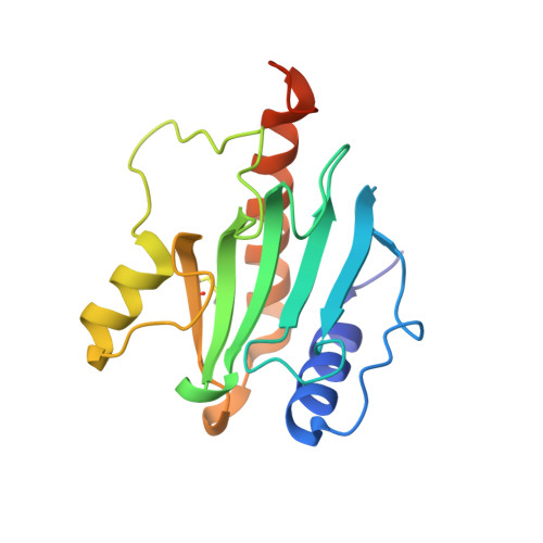
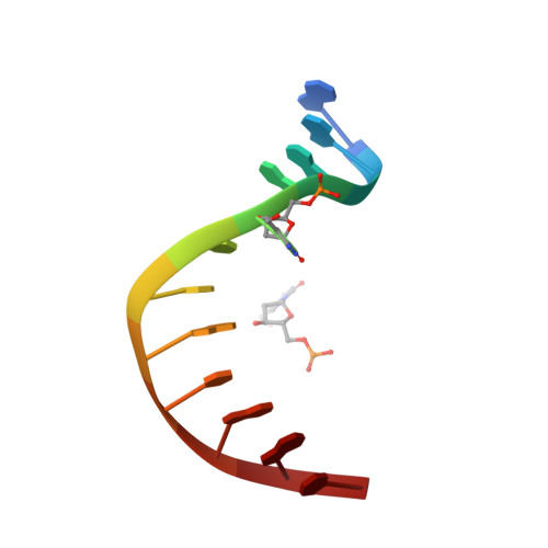
Recognition of modified cytosine variants by the DNA-binding domain of methyl-directed endonuclease McrBC.

Zagorskaite, E.Manakova, E.Sasnauskas, G.

(2018) FEBS Lett 592: 3335-3345

  • DOI: https://doi.org/10.1002/1873-3468.13244
  • Primary Citation Related Structures: 
    6GCD, 6GCE, 6GCF

  • PubMed Abstract: 

    Cytosine modifications expand the information content of genomic DNA in both eukaryotes and prokaryotes, providing means for epigenetic regulation and self versus nonself discrimination. For example, the methyl-directed restriction endonuclease, McrBC, recognizes and cuts invading bacteriophage DNA containing 5-methylcytosine (5mC), 5-hydroxymethylcytosine (5hmC), and N4-methylcytosine (4mC), leaving the unmodified host DNA intact. Here, we present cocrystal structures of McrB-N bound to DNA oligoduplexes containing 5hmC, 5-formylcytosine (5fC), and 4mC, and characterize the relative affinity of McrB-N to various cytosine variants. We find that McrB-N flips out modified bases into a protein pocket and binds cytosine derivatives in the order of descending affinity: 4mC > 5mC > 5hmC ≫ 5fC. We also show that pocket mutations alter the relative preference of McrB-N to 5mC, 5hmC, and 4mC.


  • Organizational Affiliation
    • Institute of Biotechnology, Vilnius University, Vilnius, Lithuania.

Macromolecule Content 

  • Total Structure Weight: 46.86 kDa 
  • Atom Count: 3,803 
  • Modeled Residue Count: 336 
  • Deposited Residue Count: 364 
  • Unique protein chains: 1
  • Unique nucleic acid chains: 2

Macromolecules


Find similar proteins by:|  3D Structure
Entity ID: 1
MoleculeChains  Sequence LengthOrganismDetailsImage
5-methylcytosine-specific restriction enzyme B
A, B
170Escherichia coli K-12Mutation(s): 0 
Gene Names: mcrBrglBb4346JW5871
EC: 3.1.21
UniProt
Find proteins for P15005 (Escherichia coli (strain K12))
Explore P15005 
Go to UniProtKB:  P15005
Entity Groups
Sequence Clusters30% Identity50% Identity70% Identity90% Identity95% Identity100% Identity
UniProt GroupP15005
Sequence Annotations
Expand
Reference Sequence
Find similar nucleic acids by:  Sequence
Entity ID: 2
MoleculeChains LengthOrganismImage
DNA (5'-D(*GP*AP*GP*AP*(5HC)P*CP*GP*GP*TP*AP*GP*C)-3')12synthetic construct
Sequence Annotations
Expand
Reference Sequence
Find similar nucleic acids by:  Sequence
Entity ID: 3
MoleculeChains LengthOrganismImage
DNA (5'-D(*GP*CP*TP*AP*(5HC)P*CP*GP*GP*TP*CP*TP*C)-3')12synthetic construct
Sequence Annotations
Expand
Reference Sequence

Small Molecules

Modified Residues  1 Unique
IDChains TypeFormula2D DiagramParent
CME
Query on CME
A, B
L-PEPTIDE LINKINGC5 H11 N O3 S2CYS

Experimental Data & Validation

Experimental Data

  • Method: X-RAY DIFFRACTION
  • Resolution: 1.80 Å
  • R-Value Free:  0.214 (Depositor), 0.208 (DCC) 
  • R-Value Work:  0.166 (Depositor) 
  • R-Value Observed: 0.171 (Depositor) 
Space Group: P 21 21 21
Unit Cell:
Length ( Å )Angle ( ˚ )
a = 36.071α = 90
b = 68.934β = 90
c = 143.708γ = 90
Software Package:
Software NamePurpose
PHENIXrefinement
XDSdata reduction
SCALAdata scaling
MOLREPphasing

Structure Validation

View Full Validation Report


Entry History 

& Funding Information

Deposition Data


Funding OrganizationLocationGrant Number
LithuaniaResearch Council of Lithuania, MIP-027/2012

Revision History  (Full details and data files)

  • Version 1.0: 2018-09-19
    Type: Initial release
  • Version 1.1: 2018-10-24
    Changes: Data collection, Database references
  • Version 1.2: 2024-01-17
    Changes: Data collection, Database references, Refinement description
  • Version 1.3: 2024-11-13
    Changes: Structure summary