6GBW | pdb_00006gbw

Thrombin in complex with MI2100 ((S)-N-(2-(aminomethyl)-5-chlorobenzyl)-1-((benzylsulfonyl)-L-arginyl)pyrrolidine-2-carboxamide)


Experimental Data Snapshot

  • Method: X-RAY DIFFRACTION
  • Resolution: 1.45 Å
  • R-Value Free: 
    0.165 (Depositor), 0.167 (DCC) 
  • R-Value Work: 
    0.132 (Depositor), 0.134 (DCC) 
  • R-Value Observed: 
    0.134 (Depositor) 

Starting Model: experimental
View more details

wwPDB Validation 3D Report Full Report

Validation slider image for 6GBW

Ligand Structure Quality Assessment 


This is version 1.4 of the entry. See complete history

Literature

Strategies for Late-Stage Optimization: Profiling Thermodynamics by Preorganization and Salt Bridge Shielding.

Sandner, A.Hufner-Wulsdorf, T.Heine, A.Steinmetzer, T.Klebe, G.

(2019) J Med Chem 62: 9753-9771

  • DOI: https://doi.org/10.1021/acs.jmedchem.9b01196
  • Primary Citation Related Structures: 
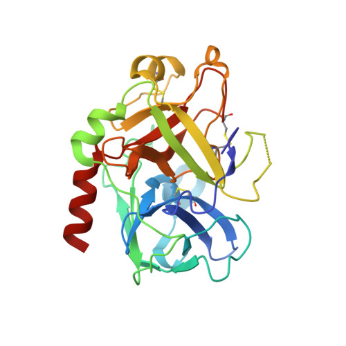
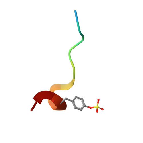
    5JFD, 5JZY, 5LCE, 5LPD, 6GBW, 6ROT

  • PubMed Abstract: 

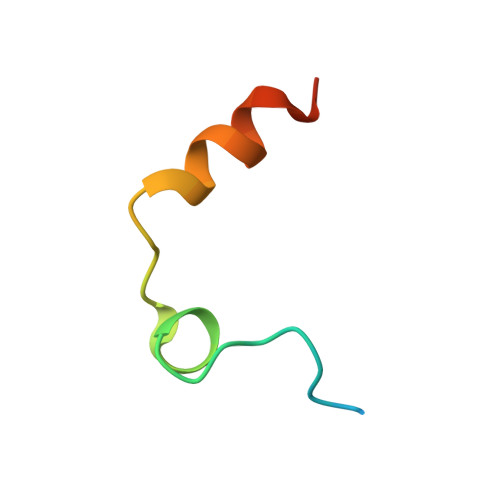
    Structural fixation of a ligand in its bioactive conformation may, due to entropic reasons, improve affinity. We present a congeneric series of thrombin ligands with a variety of functional groups triggering preorganization prior to binding. Fixation in solution and complex formation have been characterized by crystallography, isothermal titration calorimetry (ITC), and molecular dynamics (MD) simulations. First, we show why these preorganizing modifications do not affect the overall binding mode and how key interactions are preserved. Next, we demonstrate how preorganization thermodynamics can be largely dominated by enthalpy rather than entropy because of the significant population of low-energy conformations. Furthermore, a salt bridge is shielded by actively reducing its surface exposure, thus leading to an enhanced enthalpic binding profile. Our results suggest that the consideration of the ligand solution ensemble by MD simulation is necessary to predict preorganizing modifications that enhance the binding behavior of already promising binders.


  • Organizational Affiliation
    • Institut für Pharmazeutische Chemie , Philipps-Universität Marburg , Marbacher Weg 6 , 35032 Marburg , Germany.

Macromolecule Content 

  • Total Structure Weight: 36.43 kDa 
  • Atom Count: 2,761 
  • Modeled Residue Count: 289 
  • Deposited Residue Count: 307 
  • Unique protein chains: 3

Macromolecules

Find similar proteins by:|  3D Structure
Entity ID: 1
MoleculeChains  Sequence LengthOrganismDetailsImage
ProthrombinA [auth L]36Homo sapiensMutation(s): 0 
EC: 3.4.21.5
UniProt & NIH Common Fund Data Resources
Find proteins for P00734 (Homo sapiens)
Explore P00734 
Go to UniProtKB:  P00734
PHAROS:  P00734
GTEx:  ENSG00000180210 
Entity Groups
Sequence Clusters30% Identity50% Identity70% Identity90% Identity95% Identity100% Identity
UniProt GroupP00734
Sequence Annotations
Expand
Reference Sequence
Find similar proteins by:|  3D Structure
Entity ID: 2
MoleculeChains  Sequence LengthOrganismDetailsImage
ProthrombinB [auth H]259Homo sapiensMutation(s): 0 
EC: 3.4.21.5
UniProt & NIH Common Fund Data Resources
Find proteins for P00734 (Homo sapiens)
Explore P00734 
Go to UniProtKB:  P00734
PHAROS:  P00734
GTEx:  ENSG00000180210 
Entity Groups
Sequence Clusters30% Identity50% Identity70% Identity90% Identity95% Identity100% Identity
UniProt GroupP00734
Sequence Annotations
Expand
Reference Sequence
Find similar proteins by:  Sequence   |   3D Structure  
Entity ID: 3
MoleculeChains  Sequence LengthOrganismDetailsImage
Hirudin variant-2C [auth I]12Hirudo medicinalisMutation(s): 0 
UniProt
Find proteins for P09945 (Hirudo medicinalis)
Explore P09945 
Go to UniProtKB:  P09945
Entity Groups
Sequence Clusters30% Identity50% Identity70% Identity90% Identity95% Identity100% Identity
UniProt GroupP09945
Sequence Annotations
Expand
Reference Sequence

Small Molecules

Ligands 4 Unique
IDChains Name / Formula / InChI Key2D Diagram3D Interactions
EU5

Query on EU5


Download:Ideal Coordinates CCD File
I [auth H](2~{S})-~{N}-[[2-(aminomethyl)-5-chloranyl-phenyl]methyl]-1-[(2~{S})-5-carbamimidamido-2-[(phenylmethyl)sulfonylamino]pentanoyl]pyrrolidine-2-carboxamide
C26 H36 Cl N7 O4 S
MWLLGEVAEDIFRC-GOTSBHOMSA-N
NAG

Query on NAG


Download:Ideal Coordinates CCD File
D [auth H]2-acetamido-2-deoxy-beta-D-glucopyranose
C8 H15 N O6
OVRNDRQMDRJTHS-FMDGEEDCSA-N
DMS

Query on DMS


Download:Ideal Coordinates CCD File
G [auth H],
H
DIMETHYL SULFOXIDE
C2 H6 O S
IAZDPXIOMUYVGZ-UHFFFAOYSA-N
NA

Query on NA


Download:Ideal Coordinates CCD File
E [auth H],
F [auth H]
SODIUM ION
Na
FKNQFGJONOIPTF-UHFFFAOYSA-N
Modified Residues  1 Unique
IDChains TypeFormula2D DiagramParent
TYS
Query on TYS
C [auth I]L-PEPTIDE LINKINGC9 H11 N O6 STYR
Binding Affinity Annotations 
IDSourceBinding Affinity
EU5 BindingDB:  6GBW Ki: 3.5 (nM) from 1 assay(s)

Experimental Data & Validation

Experimental Data

  • Method: X-RAY DIFFRACTION
  • Resolution: 1.45 Å
  • R-Value Free:  0.165 (Depositor), 0.167 (DCC) 
  • R-Value Work:  0.132 (Depositor), 0.134 (DCC) 
  • R-Value Observed: 0.134 (Depositor) 
Space Group: C 1 2 1
Unit Cell:
Length ( Å )Angle ( ˚ )
a = 69.84α = 90
b = 71.119β = 100.58
c = 72.858γ = 90
Software Package:
Software NamePurpose
PHENIXrefinement
Cootmodel building
XDSdata scaling
PHASERphasing
XDSdata reduction

Structure Validation

View Full Validation Report


Ligand Structure Quality Assessment 


Entry History 

Deposition Data

Revision History  (Full details and data files)

  • Version 1.0: 2019-04-24
    Type: Initial release
  • Version 1.1: 2019-11-27
    Changes: Database references
  • Version 1.2: 2020-07-29
    Type: Remediation
    Reason: Carbohydrate remediation
    Changes: Data collection, Derived calculations, Structure summary
  • Version 1.3: 2024-01-17
    Changes: Data collection, Database references, Refinement description, Structure summary
  • Version 1.4: 2024-11-13
    Changes: Structure summary