6G8G

Flavonoid-responsive Regulator FrrA in complex with Genistein


Experimental Data Snapshot

  • Method: X-RAY DIFFRACTION
  • Resolution: 2.60 Å
  • R-Value Free: 0.216 
  • R-Value Work: 0.188 
  • R-Value Observed: 0.190 

Starting Model: experimental
View more details

wwPDB Validation   3D Report Full Report


Ligand Structure Quality Assessment 


This is version 1.2 of the entry. See complete history


Literature

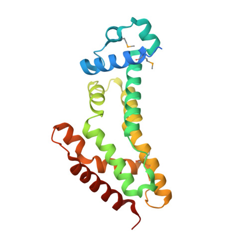
The induction mechanism of the flavonoid-responsive regulator FrrA.

Werner, N.Werten, S.Hoppen, J.Palm, G.J.Gottfert, M.Hinrichs, W.

(2021) FEBS J 

  • DOI: https://doi.org/10.1111/febs.16141
  • Primary Citation of Related Structures:  
    6G87, 6G8G, 6G8H

  • PubMed Abstract: 

    Bradyrhizobium diazoefficiens, a bacterial symbiont of soybean and other leguminous plants, enters a nodulation-promoting genetic programme in the presence of host-produced flavonoids and related signalling compounds. Here, we describe the crystal structure of an isoflavonoid-responsive regulator (FrrA) from Bradyrhizobium, as well as cocrystal structures with inducing and noninducing ligands (genistein and naringenin, respectively). The structures reveal a TetR-like fold whose DNA-binding domain is capable of adopting a range of orientations. A single molecule of either genistein or naringenin is asymmetrically bound in a central cavity of the FrrA homodimer, mainly via C-H contacts to the π-system of the ligands. Strikingly, however, the interaction does not provoke any conformational changes in the repressor. Both the flexible positioning of the DNA-binding domain and the absence of structural change upon ligand binding are corroborated by small-angle X-ray scattering (SAXS) experiments in solution. Together with a model of the promoter-bound state of FrrA our results suggest that inducers act as a wedge, preventing the DNA-binding domains from moving close enough together to interact with successive positions of the major groove of the palindromic operator.


  • Organizational Affiliation

    Institute for Biochemistry, Department Molecular Structural Biology, University of Greifswald, Germany.


Macromolecules
Find similar proteins by:  (by identity cutoff)  |  3D Structure
Entity ID: 1
MoleculeChains Sequence LengthOrganismDetailsImage
TetR/AcrR family transcriptional regulator
A, B, C, D
214Bradyrhizobium diazoefficiensMutation(s): 0 
Gene Names: CO678_15510
Entity Groups  
Sequence Clusters30% Identity50% Identity70% Identity90% Identity95% Identity100% Identity
Sequence Annotations
Expand
  • Reference Sequence
Small Molecules
Ligands 2 Unique
IDChains Name / Formula / InChI Key2D Diagram3D Interactions
GEN
Query on GEN

Download Ideal Coordinates CCD File 
G [auth B],
L [auth D]
GENISTEIN
C15 H10 O5
TZBJGXHYKVUXJN-UHFFFAOYSA-N
NHE
Query on NHE

Download Ideal Coordinates CCD File 
E [auth A]
F [auth A]
H [auth B]
I [auth B]
J [auth B]
E [auth A],
F [auth A],
H [auth B],
I [auth B],
J [auth B],
K [auth C],
M [auth D]
2-[N-CYCLOHEXYLAMINO]ETHANE SULFONIC ACID
C8 H17 N O3 S
MKWKNSIESPFAQN-UHFFFAOYSA-N
Modified Residues  1 Unique
IDChains TypeFormula2D DiagramParent
MSE
Query on MSE
A, B, C, D
L-PEPTIDE LINKINGC5 H11 N O2 SeMET
Experimental Data & Validation

Experimental Data

  • Method: X-RAY DIFFRACTION
  • Resolution: 2.60 Å
  • R-Value Free: 0.216 
  • R-Value Work: 0.188 
  • R-Value Observed: 0.190 
  • Space Group: P 43
Unit Cell:
Length ( Å )Angle ( ˚ )
a = 119.044α = 90
b = 119.044β = 90
c = 78.305γ = 90
Software Package:
Software NamePurpose
REFMACrefinement
XDSdata reduction
XDSdata scaling
PHASERphasing

Structure Validation

View Full Validation Report


Ligand Structure Quality Assessment 


Entry History 

Deposition Data

Revision History  (Full details and data files)

  • Version 1.0: 2019-04-17
    Type: Initial release
  • Version 1.1: 2021-08-18
    Changes: Database references
  • Version 1.2: 2024-01-17
    Changes: Data collection, Refinement description