6G7O | pdb_00006g7o

Crystal structure of human alkaline ceramidase 3 (ACER3) at 2.7 Angstrom resolution


Experimental Data Snapshot

  • Method: X-RAY DIFFRACTION
  • Resolution: 2.70 Å
  • R-Value Free: 
    0.270 (Depositor), 0.263 (DCC) 
  • R-Value Work: 
    0.248 (Depositor), 0.259 (DCC) 
  • R-Value Observed: 
    0.249 (Depositor) 

Starting Model: experimental
View more details

wwPDB Validation 3D Report Full Report

Validation slider image for 6G7O

Ligand Structure Quality Assessment 


This is version 1.2 of the entry. See complete history

Literature

Structure of a human intramembrane ceramidase explains enzymatic dysfunction found in leukodystrophy.

Vasiliauskaite-Brooks, I.Healey, R.D.Rochaix, P.Saint-Paul, J.Sounier, R.Grison, C.Waltrich-Augusto, T.Fortier, M.Hoh, F.Saied, E.M.Arenz, C.Basu, S.Leyrat, C.Granier, S.

(2018) Nat Commun 9: 5437-5437

  • DOI: https://doi.org/10.1038/s41467-018-07864-w
  • Primary Citation Related Structures: 
    6G7O

  • PubMed Abstract: 

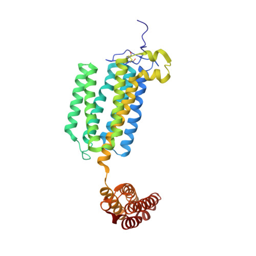
    Alkaline ceramidases (ACERs) are a class of poorly understood transmembrane enzymes controlling the homeostasis of ceramides. They are implicated in human pathophysiology, including progressive leukodystrophy, colon cancer as well as acute myeloid leukemia. We report here the crystal structure of the human ACER type 3 (ACER3). Together with computational studies, the structure reveals that ACER3 is an intramembrane enzyme with a seven transmembrane domain architecture and a catalytic Zn 2+ binding site in its core, similar to adiponectin receptors. Interestingly, we uncover a Ca 2+ binding site physically and functionally connected to the Zn 2+ providing a structural explanation for the known regulatory role of Ca 2+ on ACER3 enzymatic activity and for the loss of function in E33G-ACER3 mutant found in leukodystrophic patients.


  • Organizational Affiliation
    • IGF, University of Montpellier, CNRS, INSERM, Montpellier, 34094, France.

Macromolecule Content 

  • Total Structure Weight: 41.65 kDa 
  • Atom Count: 3,039 
  • Modeled Residue Count: 350 
  • Deposited Residue Count: 350 
  • Unique protein chains: 1

Macromolecules

Find similar proteins by:|  3D Structure
Entity ID: 1
MoleculeChains  Sequence LengthOrganismDetailsImage
Alkaline ceramidase 3,Soluble cytochrome b562350Homo sapiensEscherichia coli
This entity is chimeric
Mutation(s): 0 
Gene Names: ACER3APHCPHCAcybC
EC: 3.5.1 (PDB Primary Data), 3.5.1.23 (UniProt)
Membrane Entity: Yes 
UniProt & NIH Common Fund Data Resources
Find proteins for P0ABE7 (Escherichia coli)
Explore P0ABE7 
Go to UniProtKB:  P0ABE7
Find proteins for Q9NUN7 (Homo sapiens)
Explore Q9NUN7 
Go to UniProtKB:  Q9NUN7
PHAROS:  Q9NUN7
GTEx:  ENSG00000078124 
Entity Groups
Sequence Clusters30% Identity50% Identity70% Identity90% Identity95% Identity100% Identity
UniProt GroupsP0ABE7Q9NUN7
Sequence Annotations
Expand
Reference Sequence

Small Molecules

Ligands 6 Unique
IDChains Name / Formula / InChI Key2D Diagram3D Interactions
OLB

Query on OLB


Download:Ideal Coordinates CCD File
L [auth A](2S)-2,3-dihydroxypropyl (9Z)-octadec-9-enoate
C21 H40 O4
RZRNAYUHWVFMIP-QJRAZLAKSA-N
SO4

Query on SO4


Download:Ideal Coordinates CCD File
C [auth A]
D [auth A]
E [auth A]
F [auth A]
G [auth A]
C [auth A],
D [auth A],
E [auth A],
F [auth A],
G [auth A],
H [auth A]
SULFATE ION
O4 S
QAOWNCQODCNURD-UHFFFAOYSA-L
ZN

Query on ZN


Download:Ideal Coordinates CCD File
B [auth A]ZINC ION
Zn
PTFCDOFLOPIGGS-UHFFFAOYSA-N
CA

Query on CA


Download:Ideal Coordinates CCD File
M [auth A]CALCIUM ION
Ca
BHPQYMZQTOCNFJ-UHFFFAOYSA-N
MG

Query on MG


Download:Ideal Coordinates CCD File
N [auth A],
O [auth A]
MAGNESIUM ION
Mg
JLVVSXFLKOJNIY-UHFFFAOYSA-N
NA

Query on NA


Download:Ideal Coordinates CCD File
I [auth A],
J [auth A],
K [auth A]
SODIUM ION
Na
FKNQFGJONOIPTF-UHFFFAOYSA-N

Experimental Data & Validation

Experimental Data

  • Method: X-RAY DIFFRACTION
  • Resolution: 2.70 Å
  • R-Value Free:  0.270 (Depositor), 0.263 (DCC) 
  • R-Value Work:  0.248 (Depositor), 0.259 (DCC) 
  • R-Value Observed: 0.249 (Depositor) 
Space Group: C 2 2 21
Unit Cell:
Length ( Å )Angle ( ˚ )
a = 60.88α = 90
b = 68.83β = 90
c = 257.52γ = 90
Software Package:
Software NamePurpose
BUSTERrefinement
XDSdata reduction
Aimlessdata scaling
PHASERphasing
PDB_EXTRACTdata extraction

Structure Validation

View Full Validation Report


Ligand Structure Quality Assessment 


Entry History 

& Funding Information

Deposition Data


Funding OrganizationLocationGrant Number
European CommissionFrance647687

Revision History  (Full details and data files)

  • Version 1.0: 2019-01-02
    Type: Initial release
  • Version 1.1: 2024-01-17
    Changes: Advisory, Data collection, Database references, Derived calculations, Refinement description
  • Version 1.2: 2024-11-06
    Changes: Structure summary