


Direct visible light activation of a surface cysteine-engineered [NiFe]-hydrogenase by silver nanoclusters
Zhang, L., Beaton, S.E., Carr, S.B., Armstrong, F.A.(2019) Energy Environ Sci
Experimental Data Snapshot
Starting Model: experimental
View more details
wwPDB Validation 3D Report Full Report

(2019) Energy Environ Sci
Entity ID: 1 | |||||
|---|---|---|---|---|---|
| Molecule | Chains | Sequence Length | Organism | Details | Image |
| Hydrogenase-2 small chain | A [auth S], C [auth T] | 304 | Escherichia coli K-12 | Mutation(s): 0 EC: 1.12.99.6 | 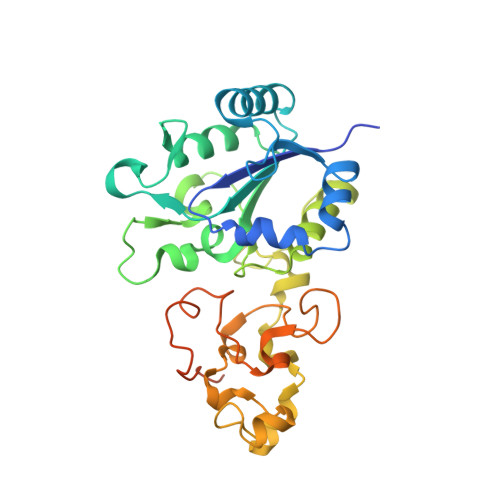 |
UniProt | |||||
Entity Groups | |||||
| Sequence Clusters | 30% Identity50% Identity70% Identity90% Identity95% Identity100% Identity | ||||
| UniProt Group | P69741 | ||||
Sequence AnnotationsExpand | |||||
Reference Sequence | |||||
Entity ID: 2 | |||||
|---|---|---|---|---|---|
| Molecule | Chains | Sequence Length | Organism | Details | Image |
| Hydrogenase-2 large chain | B [auth L], D [auth M] | 567 | Escherichia coli K-12 | Mutation(s): 3 EC: 1.12.99.6 | 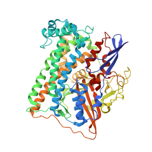 |
UniProt | |||||
Entity Groups | |||||
| Sequence Clusters | 30% Identity50% Identity70% Identity90% Identity95% Identity100% Identity | ||||
| UniProt Group | P0ACE0 | ||||
Sequence AnnotationsExpand | |||||
Reference Sequence | |||||
| Ligands 5 Unique | |||||
|---|---|---|---|---|---|
| ID | Chains | Name / Formula / InChI Key | 2D Diagram | 3D Interactions | |
| SF4 Download:Ideal Coordinates CCD File | E [auth S], G [auth S], L [auth T], N [auth T] | IRON/SULFUR CLUSTER Fe4 S4 LJBDFODJNLIPKO-UHFFFAOYSA-N |  | ||
| F3S Download:Ideal Coordinates CCD File | F [auth S], M [auth T] | FE3-S4 CLUSTER Fe3 S4 FCXHZBQOKRZXKS-UHFFFAOYSA-N |  | ||
| FCO Download:Ideal Coordinates CCD File | H [auth L], O [auth M] | CARBONMONOXIDE-(DICYANO) IRON C3 Fe N2 O VBQUCMTXYFMTTE-UHFFFAOYSA-N |  | ||
| NI Download:Ideal Coordinates CCD File | I [auth L], P [auth M] | NICKEL (II) ION Ni VEQPNABPJHWNSG-UHFFFAOYSA-N |  | ||
| MG Download:Ideal Coordinates CCD File | J [auth L], K [auth L], Q [auth M], R [auth M] | MAGNESIUM ION Mg JLVVSXFLKOJNIY-UHFFFAOYSA-N |  | ||
| Length ( Å ) | Angle ( ˚ ) |
|---|---|
| a = 100.237 | α = 90 |
| b = 100.986 | β = 90 |
| c = 169.425 | γ = 90 |
| Software Name | Purpose |
|---|---|
| REFMAC | refinement |
| XDS | data reduction |
| Aimless | data scaling |
| PHASER | phasing |
| Funding Organization | Location | Grant Number |
|---|---|---|
| Biotechnology and Biological Sciences Research Council | United Kingdom | BB/N006321/1 |


