


Mutational and structural studies uncover crucial amino acids determining activity and stability of 17beta-HSD14
Badran, M., Bertoletti, N., Keils, A., Heine, A., Klebe, G., Marchais-Oberwinkler, S.(2019) J Steroid Biochem Mol Biol
Experimental Data Snapshot
Starting Model: experimental
View more details
(2019) J Steroid Biochem Mol Biol
Entity ID: 1 | |||||
|---|---|---|---|---|---|
| Molecule | Chains | Sequence Length | Organism | Details | Image |
| 17-beta-hydroxysteroid dehydrogenase 14 | 270 | Homo sapiens | Mutation(s): 1 Gene Names: HSD17B14, DHRS10, SDR3, SDR47C1, UNQ502/PRO474 EC: 1.1.1 (PDB Primary Data), 1.1.1.122 (UniProt) | 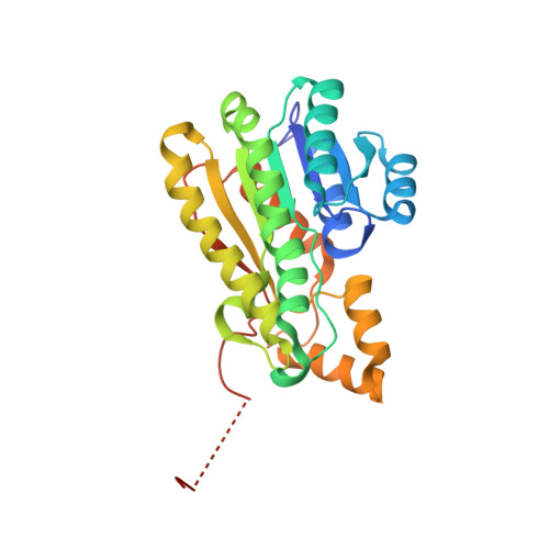 | |
UniProt & NIH Common Fund Data Resources | |||||
PHAROS: Q9BPX1 GTEx: ENSG00000087076 | |||||
Entity Groups | |||||
| Sequence Clusters | 30% Identity50% Identity70% Identity90% Identity95% Identity100% Identity | ||||
| UniProt Group | Q9BPX1 | ||||
Sequence AnnotationsExpand | |||||
Reference Sequence | |||||
| Ligands 5 Unique | |||||
|---|---|---|---|---|---|
| ID | Chains | Name / Formula / InChI Key | 2D Diagram | 3D Interactions | |
| NAD Download:Ideal Coordinates CCD File | B [auth A] | NICOTINAMIDE-ADENINE-DINUCLEOTIDE C21 H27 N7 O14 P2 BAWFJGJZGIEFAR-NNYOXOHSSA-N |  | ||
| F45 Download:Ideal Coordinates CCD File | E [auth A] | [6-(3,4-dihydroxyphenyl)pyridin-2-yl](4-fluoro-3-hydroxyphenyl)methanone C18 H12 F N O4 USZFAGSFWNNZLX-UHFFFAOYSA-N |  | ||
| BGC Download:Ideal Coordinates CCD File | D [auth A] | beta-D-glucopyranose C6 H12 O6 WQZGKKKJIJFFOK-VFUOTHLCSA-N |  | ||
| DMS Download:Ideal Coordinates CCD File | F [auth A], G [auth A] | DIMETHYL SULFOXIDE C2 H6 O S IAZDPXIOMUYVGZ-UHFFFAOYSA-N |  | ||
| NA Download:Ideal Coordinates CCD File | C [auth A] | SODIUM ION Na FKNQFGJONOIPTF-UHFFFAOYSA-N |  | ||
| Length ( Å ) | Angle ( ˚ ) |
|---|---|
| a = 91.423 | α = 90 |
| b = 91.423 | β = 90 |
| c = 132.141 | γ = 90 |
| Software Name | Purpose |
|---|---|
| PHENIX | refinement |
| XDS | data reduction |
| XDS | data scaling |
| PHASER | phasing |


