6G37

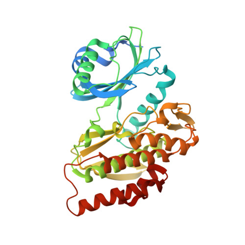
Crystal structure of haspin in complex with 5-fluorotubercidin


Experimental Data Snapshot

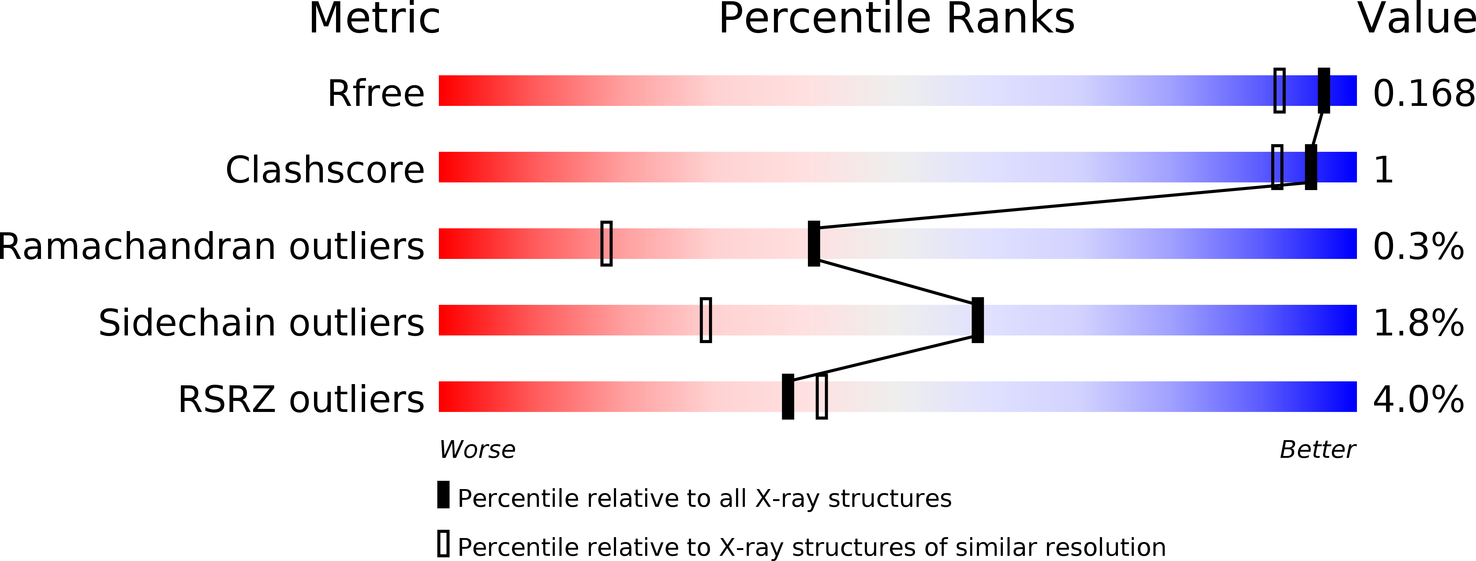
  • Method: X-RAY DIFFRACTION
  • Resolution: 1.48 Å
  • R-Value Free: 0.167 
  • R-Value Work: 0.148 
  • R-Value Observed: 0.149 

Starting Model: experimental
View more details

wwPDB Validation   3D Report Full Report


Ligand Structure Quality Assessment 


This is version 1.3 of the entry. See complete history


Literature

Halogen-Aromatic pi Interactions Modulate Inhibitor Residence Times.

Heroven, C.Georgi, V.Ganotra, G.K.Brennan, P.Wolfreys, F.Wade, R.C.Fernandez-Montalvan, A.E.Chaikuad, A.Knapp, S.

(2018) Angew Chem Int Ed Engl 57: 7220-7224

  • DOI: https://doi.org/10.1002/anie.201801666
  • Primary Citation of Related Structures:  
    6G33, 6G34, 6G35, 6G36, 6G37, 6G38, 6G39, 6G3A

  • PubMed Abstract: 

    Prolonged drug residence times may result in longer-lasting drug efficacy, improved pharmacodynamic properties, and "kinetic selectivity" over off-targets with high drug dissociation rates. However, few strategies have been elaborated to rationally modulate drug residence time and thereby to integrate this key property into the drug development process. Herein, we show that the interaction between a halogen moiety on an inhibitor and an aromatic residue in the target protein can significantly increase inhibitor residence time. By using the interaction of the serine/threonine kinase haspin with 5-iodotubercidin (5-iTU) derivatives as a model for an archetypal active-state (type I) kinase-inhibitor binding mode, we demonstrate that inhibitor residence times markedly increase with the size and polarizability of the halogen atom. The halogen-aromatic π interactions in the haspin-inhibitor complexes were characterized by means of kinetic, thermodynamic, and structural measurements along with binding-energy calculations.


  • Organizational Affiliation

    Nuffield Department of Clinical Medicine, Structural Genomics Consortium, University of Oxford, Oxford, OX3 7DQ, UK.


Macromolecules
Find similar proteins by:  (by identity cutoff)  |  3D Structure
Entity ID: 1
MoleculeChains Sequence LengthOrganismDetailsImage
Serine/threonine-protein kinase haspin357Homo sapiensMutation(s): 0 
Gene Names: HASPINGSG2
EC: 2.7.11.1
UniProt & NIH Common Fund Data Resources
Find proteins for Q8TF76 (Homo sapiens)
Explore Q8TF76 
Go to UniProtKB:  Q8TF76
PHAROS:  Q8TF76
GTEx:  ENSG00000177602 
Entity Groups  
Sequence Clusters30% Identity50% Identity70% Identity90% Identity95% Identity100% Identity
UniProt GroupQ8TF76
Sequence Annotations
Expand
  • Reference Sequence
Small Molecules
Ligands 2 Unique
IDChains Name / Formula / InChI Key2D Diagram3D Interactions
FTU
Query on FTU

Download Ideal Coordinates CCD File 
C [auth A]5-fluorotubercidin
C11 H13 F N4 O4
KRRZKBNCONNKPI-IOSLPCCCSA-N
PO4
Query on PO4

Download Ideal Coordinates CCD File 
B [auth A]PHOSPHATE ION
O4 P
NBIIXXVUZAFLBC-UHFFFAOYSA-K
Experimental Data & Validation

Experimental Data

  • Method: X-RAY DIFFRACTION
  • Resolution: 1.48 Å
  • R-Value Free: 0.167 
  • R-Value Work: 0.148 
  • R-Value Observed: 0.149 
  • Space Group: P 21 21 21
Unit Cell:
Length ( Å )Angle ( ˚ )
a = 78.711α = 90
b = 78.89β = 90
c = 79.63γ = 90
Software Package:
Software NamePurpose
REFMACrefinement
iMOSFLMdata reduction
Aimlessdata scaling
PHASERphasing

Structure Validation

View Full Validation Report


Ligand Structure Quality Assessment 


Entry History 

Deposition Data

Revision History  (Full details and data files)

  • Version 1.0: 2018-04-18
    Type: Initial release
  • Version 1.1: 2018-05-16
    Changes: Data collection, Database references
  • Version 1.2: 2018-06-13
    Changes: Data collection, Database references
  • Version 1.3: 2024-01-17
    Changes: Data collection, Database references, Refinement description