6FYQ

The crystal structure of a new transaminase from the marine bacterium Virgibacillus


Experimental Data Snapshot

  • Method: X-RAY DIFFRACTION
  • Resolution: 2.00 Å
  • R-Value Free: 0.219 
  • R-Value Work: 0.177 
  • R-Value Observed: 0.179 

Starting Model: experimental
View more details

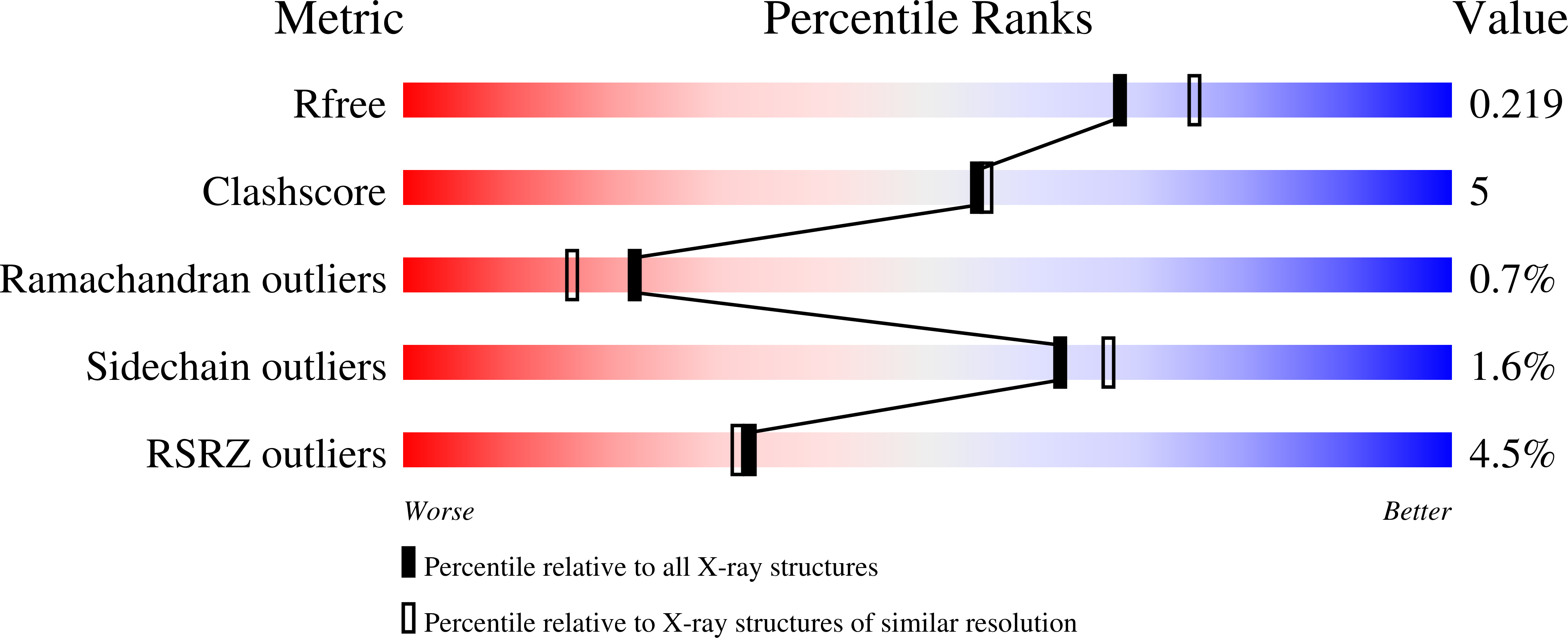
wwPDB Validation   3D Report Full Report


Ligand Structure Quality Assessment 


This is version 1.1 of the entry. See complete history


Literature

Strategic single point mutation yields a solvent- and salt-stable transaminase from Virgibacillus sp. in soluble form.

Guidi, B.Planchestainer, M.Contente, M.L.Laurenzi, T.Eberini, I.Gourlay, L.J.Romano, D.Paradisi, F.Molinari, F.

(2018) Sci Rep 8: 16441-16441

  • DOI: https://doi.org/10.1038/s41598-018-34434-3
  • Primary Citation of Related Structures:  
    6FYQ

  • PubMed Abstract: 

    A new transaminase (VbTA) was identified from the genome of the halotolerant marine bacterium Virgibacillus 21D. Following heterologous expression in Escherichia coli, it was located entirely in the insoluble fraction. After a single mutation, identified via sequence homology analyses, the VbTA T16F mutant was successfully expressed in soluble form and characterised. VbTA T16F showed high stability towards polar organic solvents and salt exposure, accepting mainly hydrophobic aromatic amine and carbonyl substrates. The 2.0 Å resolution crystal structure of VbTA T16F is here reported, and together with computational calculations, revealed that this mutation is crucial for correct dimerisation and thus correct folding, leading to soluble protein expression.


  • Organizational Affiliation

    Department of Food, Environmental and Nutritional Sciences (DeFENS), University of Milan, Milan, Italy.


Macromolecules
Find similar proteins by:  (by identity cutoff)  |  3D Structure
Entity ID: 1
MoleculeChains Sequence LengthOrganismDetailsImage
amine transaminase484Virgibacillus pantothenticusMutation(s): 0 
EC: 2.6.1.18
UniProt
Find proteins for A0A4P1LYG1 (Virgibacillus pantothenticus)
Explore A0A4P1LYG1 
Go to UniProtKB:  A0A4P1LYG1
Entity Groups  
Sequence Clusters30% Identity50% Identity70% Identity90% Identity95% Identity100% Identity
UniProt GroupA0A4P1LYG1
Sequence Annotations
Expand
  • Reference Sequence
Experimental Data & Validation

Experimental Data

  • Method: X-RAY DIFFRACTION
  • Resolution: 2.00 Å
  • R-Value Free: 0.219 
  • R-Value Work: 0.177 
  • R-Value Observed: 0.179 
  • Space Group: P 32 2 1
Unit Cell:
Length ( Å )Angle ( ˚ )
a = 100.886α = 90
b = 100.886β = 90
c = 97.411γ = 120
Software Package:
Software NamePurpose
XDSdata reduction
SCALAdata scaling
MOLREPphasing
PHENIXrefinement
PDB_EXTRACTdata extraction

Structure Validation

View Full Validation Report


Ligand Structure Quality Assessment 


Entry History 

Deposition Data

Revision History  (Full details and data files)

  • Version 1.0: 2018-12-12
    Type: Initial release
  • Version 1.1: 2024-01-17
    Changes: Advisory, Data collection, Database references, Refinement description