6FWZ | pdb_00006fwz

Crystal structure of human UDP-N-acetylglucosamine-dolichyl-phosphate N-acetylglucosaminephosphotransferase (DPAGT1) (V264G mutant) in complex with UDP-GlcNAc


Experimental Data Snapshot

  • Method: X-RAY DIFFRACTION
  • Resolution: 3.10 Å
  • R-Value Free: 
    0.236 (Depositor), 0.235 (DCC) 
  • R-Value Work: 
    0.223 (Depositor), 0.236 (DCC) 
  • R-Value Observed: 
    0.223 (Depositor) 

Starting Model: experimental
View more details

wwPDB Validation 3D Report Full Report

Validation slider image for 6FWZ

Ligand Structure Quality Assessment 


This is version 1.2 of the entry. See complete history

Literature

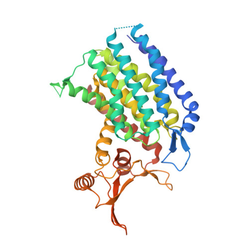
Structures of DPAGT1 Explain Glycosylation Disease Mechanisms and Advance TB Antibiotic Design.

Dong, Y.Y.Wang, H.Pike, A.C.W.Cochrane, S.A.Hamedzadeh, S.Wyszynski, F.J.Bushell, S.R.Royer, S.F.Widdick, D.A.Sajid, A.Boshoff, H.I.Park, Y.Lucas, R.Liu, W.M.Lee, S.S.Machida, T.Minall, L.Mehmood, S.Belaya, K.Liu, W.W.Chu, A.Shrestha, L.Mukhopadhyay, S.M.M.Strain-Damerell, C.Chalk, R.Burgess-Brown, N.A.Bibb, M.J.Barry Iii, C.E.Robinson, C.V.Beeson, D.Davis, B.G.Carpenter, E.P.

(2018) Cell 175: 1045-1058.e16

  • DOI: https://doi.org/10.1016/j.cell.2018.10.037
  • Primary Citation Related Structures: 
    5LEV, 5O5E, 6FM9, 6FWZ

  • PubMed Abstract: 

    Protein N-glycosylation is a widespread post-translational modification. The first committed step in this process is catalysed by dolichyl-phosphate N-acetylglucosamine-phosphotransferase DPAGT1 (GPT/E.C. 2.7.8.15). Missense DPAGT1 variants cause congenital myasthenic syndrome and disorders of glycosylation. In addition, naturally-occurring bactericidal nucleoside analogues such as tunicamycin are toxic to eukaryotes due to DPAGT1 inhibition, preventing their clinical use. Our structures of DPAGT1 with the substrate UDP-GlcNAc and tunicamycin reveal substrate binding modes, suggest a mechanism of catalysis, provide an understanding of how mutations modulate activity (thus causing disease) and allow design of non-toxic "lipid-altered" tunicamycins. The structure-tuned activity of these analogues against several bacterial targets allowed the design of potent antibiotics for Mycobacterium tuberculosis, enabling treatment in vitro, in cellulo and in vivo, providing a promising new class of antimicrobial drug.


  • Organizational Affiliation
    • Structural Genomics Consortium, University of Oxford, Oxford, OX3 7DQ, UK.

Macromolecule Content 

  • Total Structure Weight: 47.55 kDa 
  • Atom Count: 2,998 
  • Modeled Residue Count: 373 
  • Deposited Residue Count: 409 
  • Unique protein chains: 1

Macromolecules

Find similar proteins by:|  3D Structure
Entity ID: 1
MoleculeChains  Sequence LengthOrganismDetailsImage
UDP-N-acetylglucosamine--dolichyl-phosphate N-acetylglucosaminephosphotransferase409Homo sapiensMutation(s): 1 
Gene Names: DPAGT1DPAGT2
EC: 2.7.8.15
Membrane Entity: Yes 
UniProt & NIH Common Fund Data Resources
Find proteins for Q9H3H5 (Homo sapiens)
Explore Q9H3H5 
Go to UniProtKB:  Q9H3H5
PHAROS:  Q9H3H5
GTEx:  ENSG00000172269 
Entity Groups
Sequence Clusters30% Identity50% Identity70% Identity90% Identity95% Identity100% Identity
UniProt GroupQ9H3H5
Sequence Annotations
Expand
Reference Sequence

Small Molecules

Ligands 4 Unique
IDChains Name / Formula / InChI Key2D Diagram3D Interactions
P6L

Query on P6L


Download:Ideal Coordinates CCD File
E [auth A](2S)-3-{[{[(2S)-2,3-DIHYDROXYPROPYL]OXY}(HYDROXY)PHOSPHORYL]OXY}-2-[(6E)-HEXADEC-6-ENOYLOXY]PROPYL (8E)-OCTADEC-8-ENOATE
C40 H75 O10 P
WLVNRDXLPAAELE-GDRYEXQRSA-N
UD1

Query on UD1


Download:Ideal Coordinates CCD File
C [auth A]URIDINE-DIPHOSPHATE-N-ACETYLGLUCOSAMINE
C17 H27 N3 O17 P2
LFTYTUAZOPRMMI-CFRASDGPSA-N
MG

Query on MG


Download:Ideal Coordinates CCD File
B [auth A]MAGNESIUM ION
Mg
JLVVSXFLKOJNIY-UHFFFAOYSA-N
UNL

Query on UNL


Download:Ideal Coordinates CCD File
D [auth A]Unknown ligand
JLVVSXFLKOJNIY-UHFFFAOYSA-N

Experimental Data & Validation

Experimental Data

  • Method: X-RAY DIFFRACTION
  • Resolution: 3.10 Å
  • R-Value Free:  0.236 (Depositor), 0.235 (DCC) 
  • R-Value Work:  0.223 (Depositor), 0.236 (DCC) 
  • R-Value Observed: 0.223 (Depositor) 
Space Group: P 65 2 2
Unit Cell:
Length ( Å )Angle ( ˚ )
a = 102.459α = 90
b = 102.459β = 90
c = 238.209γ = 120
Software Package:
Software NamePurpose
REFMACrefinement
XDSdata reduction
Aimlessdata scaling
PHASERphasing

Structure Validation

View Full Validation Report


Ligand Structure Quality Assessment 


Entry History 

& Funding Information

Deposition Data


Funding OrganizationLocationGrant Number
Wellcome TrustUnited Kingdom106169/Z/14/Z

Revision History  (Full details and data files)

  • Version 1.0: 2018-07-25
    Type: Initial release
  • Version 1.1: 2019-11-20
    Changes: Database references
  • Version 1.2: 2024-01-17
    Changes: Data collection, Database references, Derived calculations, Refinement description