6FV4 | pdb_00006fv4

The structure of N-acetyl-D-glucosamine-6-phosphate deacetylase D267A mutant from Mycobacterium smegmatis in complex with N-acetyl-D-glucosamine-6-phosphate


Experimental Data Snapshot

  • Method: X-RAY DIFFRACTION
  • Resolution: 1.97 Å
  • R-Value Free: 
    0.235 (Depositor), 0.237 (DCC) 
  • R-Value Work: 
    0.184 (Depositor), 0.186 (DCC) 
  • R-Value Observed: 
    0.186 (Depositor) 

Starting Model: experimental
View more details

wwPDB Validation 3D Report Full Report

Validation slider image for 6FV4

Ligand Structure Quality Assessment 


This is version 2.1 of the entry. See complete history

Literature

Structural and functional determination of homologs of theMycobacterium tuberculosis N-acetylglucosamine-6-phosphate deacetylase (NagA).

Ahangar, M.S.Furze, C.M.Guy, C.S.Cooper, C.Maskew, K.S.Graham, B.Cameron, A.D.Fullam, E.

(2018) J Biological Chem 293: 9770-9783

  • DOI: https://doi.org/10.1074/jbc.RA118.002597
  • Primary Citation Related Structures: 
    6FV3, 6FV4

  • PubMed Abstract: 

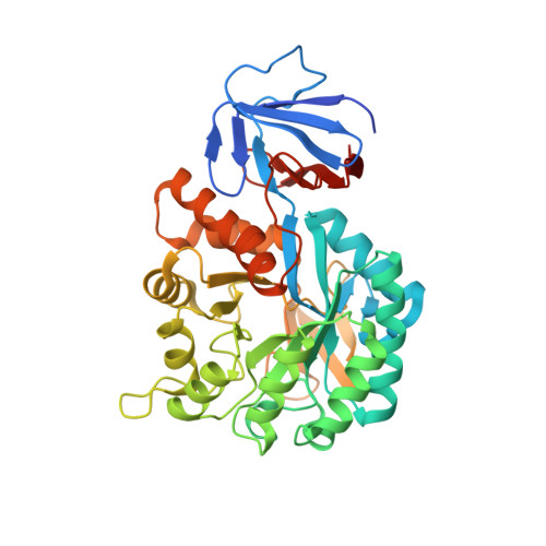
    The Mycobacterium tuberculosis ( Mtb ) pathogen encodes a GlcNAc-6-phosphate deacetylase enzyme, NagA (Rv3332), that belongs to the amidohydrolase superfamily. NagA enzymes catalyze the deacetylation of GlcNAc-6-phosphate (GlcNAc6P) to glucosamine-6-phosphate (GlcN6P). NagA is a potential antitubercular drug target because it represents the key enzymatic step in the generation of essential amino-sugar precursors required for Mtb cell wall biosynthesis and also influences recycling of cell wall peptidoglycan fragments. Here, we report the structural and functional characterization of NagA from Mycobacterium smegmatis (MSNagA) and Mycobacterium marinum (MMNagA), close relatives of Mtb Using a combination of X-ray crystallography, site-directed mutagenesis, and biochemical and biophysical assays, we show that these mycobacterial NagA enzymes are selective for GlcNAc6P. Site-directed mutagenesis studies revealed crucial roles of conserved residues in the active site that underpin stereoselective recognition, binding, and catalysis of substrates. Moreover, we report the crystal structure of MSNagA in both ligand-free form and in complex with the GlcNAc6P substrate at 2.6 and 2.0 Å resolutions, respectively. The GlcNAc6P complex structure disclosed the precise mode of GlcNAc6P binding and the structural framework of the active site, including two divalent metals located in the α/β binuclear site. Furthermore, we observed a cysteine residue located on a flexible loop region that occludes the active site. This cysteine is unique to mycobacteria and may represent a unique subsite for targeting mycobacterial NagA enzymes. Our results provide critical insights into the structural and mechanistic properties of mycobacterial NagA enzymes having an essential role in amino-sugar and nucleotide metabolism in mycobacteria.


  • Organizational Affiliation
    • From the School of Life Sciences and.

Macromolecule Content 

  • Total Structure Weight: 83.65 kDa 
  • Atom Count: 6,075 
  • Modeled Residue Count: 766 
  • Deposited Residue Count: 802 
  • Unique protein chains: 1

Macromolecules

Find similar proteins by:|  3D Structure
Entity ID: 1
MoleculeChains  Sequence LengthOrganismDetailsImage
N-acetylglucosamine-6-phosphate deacetylase
A, B
401Mycolicibacterium smegmatis MC2 155Mutation(s): 1 
Gene Names: nagAMSMEG_2119
EC: 3.5.1.25
UniProt
Find proteins for A0QU89 (Mycolicibacterium smegmatis (strain ATCC 700084 / mc(2)155))
Explore A0QU89 
Go to UniProtKB:  A0QU89
Entity Groups
Sequence Clusters30% Identity50% Identity70% Identity90% Identity95% Identity100% Identity
UniProt GroupA0QU89
Sequence Annotations
Expand
Reference Sequence

Small Molecules

Ligands 5 Unique
IDChains Name / Formula / InChI Key2D Diagram3D Interactions
16G

Query on 16G


Download:Ideal Coordinates CCD File
D [auth A]2-acetamido-2-deoxy-6-O-phosphono-alpha-D-glucopyranose
C8 H16 N O9 P
BRGMHAYQAZFZDJ-PVFLNQBWSA-N
DTT

Query on DTT


Download:Ideal Coordinates CCD File
L [auth B]2,3-DIHYDROXY-1,4-DITHIOBUTANE
C4 H10 O2 S2
VHJLVAABSRFDPM-IMJSIDKUSA-N
CD

Query on CD


Download:Ideal Coordinates CCD File
E [auth A],
F [auth A],
I [auth B],
J [auth B]
CADMIUM ION
Cd
WLZRMCYVCSSEQC-UHFFFAOYSA-N
ZN

Query on ZN


Download:Ideal Coordinates CCD File
C [auth A],
H [auth B]
ZINC ION
Zn
PTFCDOFLOPIGGS-UHFFFAOYSA-N
CL

Query on CL


Download:Ideal Coordinates CCD File
G [auth A],
K [auth B]
CHLORIDE ION
Cl
VEXZGXHMUGYJMC-UHFFFAOYSA-M

Experimental Data & Validation

Experimental Data

  • Method: X-RAY DIFFRACTION
  • Resolution: 1.97 Å
  • R-Value Free:  0.235 (Depositor), 0.237 (DCC) 
  • R-Value Work:  0.184 (Depositor), 0.186 (DCC) 
  • R-Value Observed: 0.186 (Depositor) 
Space Group: C 1 2 1
Unit Cell:
Length ( Å )Angle ( ˚ )
a = 164.766α = 90
b = 54.437β = 123.22
c = 110.262γ = 90
Software Package:
Software NamePurpose
PHENIXrefinement
XDSdata reduction
XDSdata scaling
PHASERphasing
Cootmodel building

Structure Validation

View Full Validation Report


Ligand Structure Quality Assessment 


Entry History 

& Funding Information

Deposition Data


Funding OrganizationLocationGrant Number
Wellcome TrustUnited Kingdom201442/Z/16/A
Wellcome Trust and Royal SocietyUnited Kingdom104193/Z/14/Z
Biotechnology and Biological Sciences Research CouncilUnited KingdomBB/M01116X/1
Biotechnology and Biological Sciences Research CouncilUnited KingdomBB/M017982/1
Royal SocietyUnited KingdomRG120405
Wellcome TrustUnited Kingdom105627/Z/14/Z

Revision History  (Full details and data files)

  • Version 1.0: 2018-05-16
    Type: Initial release
  • Version 1.1: 2018-07-04
    Changes: Data collection, Database references
  • Version 2.0: 2020-07-29
    Type: Remediation
    Reason: Carbohydrate remediation
    Changes: Atomic model, Data collection, Derived calculations, Structure summary
  • Version 2.1: 2024-01-17
    Changes: Data collection, Database references, Refinement description, Structure summary