Megahertz serial crystallography.
Wiedorn, M.O., Oberthur, D., Bean, R., Schubert, R., Werner, N., Abbey, B., Aepfelbacher, M., Adriano, L., Allahgholi, A., Al-Qudami, N., Andreasson, J., Aplin, S., Awel, S., Ayyer, K., Bajt, S., Barak, I., Bari, S., Bielecki, J., Botha, S., Boukhelef, D., Brehm, W., Brockhauser, S., Cheviakov, I., Coleman, M.A., Cruz-Mazo, F., Danilevski, C., Darmanin, C., Doak, R.B., Domaracky, M., Dorner, K., Du, Y., Fangohr, H., Fleckenstein, H., Frank, M., Fromme, P., Ganan-Calvo, A.M., Gevorkov, Y., Giewekemeyer, K., Ginn, H.M., Graafsma, H., Graceffa, R., Greiffenberg, D., Gumprecht, L., Gottlicher, P., Hajdu, J., Hauf, S., Heymann, M., Holmes, S., Horke, D.A., Hunter, M.S., Imlau, S., Kaukher, A., Kim, Y., Klyuev, A., Knoska, J., Kobe, B., Kuhn, M., Kupitz, C., Kupper, J., Lahey-Rudolph, J.M., Laurus, T., Le Cong, K., Letrun, R., Xavier, P.L., Maia, L., Maia, F.R.N.C., Mariani, V., Messerschmidt, M., Metz, M., Mezza, D., Michelat, T., Mills, G., Monteiro, D.C.F., Morgan, A., Muhlig, K., Munke, A., Munnich, A., Nette, J., Nugent, K.A., Nuguid, T., Orville, A.M., Pandey, S., Pena, G., Villanueva-Perez, P., Poehlsen, J., Previtali, G., Redecke, L., Riekehr, W.M., Rohde, H., Round, A., Safenreiter, T., Sarrou, I., Sato, T., Schmidt, M., Schmitt, B., Schonherr, R., Schulz, J., Sellberg, J.A., Seibert, M.M., Seuring, C., Shelby, M.L., Shoeman, R.L., Sikorski, M., Silenzi, A., Stan, C.A., Shi, X., Stern, S., Sztuk-Dambietz, J., Szuba, J., Tolstikova, A., Trebbin, M., Trunk, U., Vagovic, P., Ve, T., Weinhausen, B., White, T.A., Wrona, K., Xu, C., Yefanov, O., Zatsepin, N., Zhang, J., Perbandt, M., Mancuso, A.P., Betzel, C., Chapman, H., Barty, A.(2018) Nat Commun 9: 4025-4025
- PubMed: 30279492 Search on PubMedSearch on PubMed Central
- DOI: https://doi.org/10.1038/s41467-018-06156-7
- Primary Citation Related Structures:
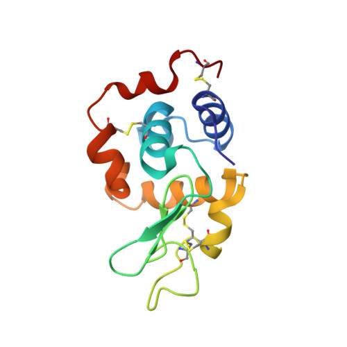
6FTR, 6GTH - PubMed Abstract:
The new European X-ray Free-Electron Laser is the first X-ray free-electron laser capable of delivering X-ray pulses with a megahertz inter-pulse spacing, more than four orders of magnitude higher than previously possible. However, to date, it has been unclear whether it would indeed be possible to measure high-quality diffraction data at megahertz pulse repetition rates. Here, we show that high-quality structures can indeed be obtained using currently available operating conditions at the European XFEL. We present two complete data sets, one from the well-known model system lysozyme and the other from a so far unknown complex of a β-lactamase from K. pneumoniae involved in antibiotic resistance. This result opens up megahertz serial femtosecond crystallography (SFX) as a tool for reliable structure determination, substrate screening and the efficient measurement of the evolution and dynamics of molecular structures using megahertz repetition rate pulses available at this new class of X-ray laser source.
- Center for Free-Electron Laser Science, Deutsches Elektronen-Synchrotron DESY, Notkestrasse 85, 22607, Hamburg, Germany.
Organizational Affiliation: