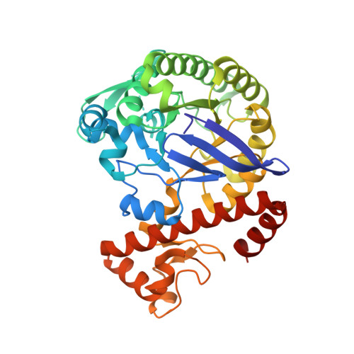
6FSO

Crystal Structure of TGT in complex with methyl({[5-(pyridin-3-yloxy)furan-2-yl]methyl})amine


Experimental Data Snapshot

  • Method: X-RAY DIFFRACTION
  • Resolution: 1.45 Å
  • R-Value Free: 0.186 
  • R-Value Work: 0.166 
  • R-Value Observed: 0.167 

Starting Model: experimental
View more details

wwPDB Validation   3D Report Full Report


Ligand Structure Quality Assessment 


This is version 2.3 of the entry. See complete history


Literature

Fragments as Novel Starting Points for tRNA-Guanine Transglycosylase Inhibitors Found by Alternative Screening Strategies.

Hassaan, E.Eriksson, P.O.Geschwindner, S.Heine, A.Klebe, G.

(2020) ChemMedChem 15: 324-337

  • DOI: https://doi.org/10.1002/cmdc.201900604
  • Primary Citation of Related Structures:  
    5N6F, 5SW3, 5UTI, 5UTJ, 5V3C, 6FSO

  • PubMed Abstract: 

    Crystallography provides structural information crucial for fragment optimization, however several criteria must be met to screen directly on protein crystals as soakable, well-diffracting specimen must be available. We screened a 96-fragment library against the tRNA-modifying enzyme TGT using crystallography. Eight hits, some with surprising binding poses, were detected. However, the amount of data collection, reduction and refinement is assumed substantial. Therefore, having a reliable cascade of fast and cost-efficient methods available for pre-screening before embarking to elaborate crystallographic screening appears beneficial. This allows filtering of compounds to the most promising hits, available to rapidly progress from hit-to-lead. But how to ensure that this workflow is reliable? To answer this question, we also applied SPR and NMR to the same screening sample to study whether identical hits are retrieved. Upon hit-list comparisons, crystallography shows with NMR and SPR, only one overlapping hit and all three methods shared no common hits. This questions a cascade-type screening protocol at least in the current example. Compared to crystallography, SPR and NMR detected higher percentages of non-active-site binders suggesting the importance of running reporter ligand-based competitive screens in SPR and NMR, a requirement not needed in crystallography. Although not specific, NMR proved a more sensitive method relative to SPR and crystallography, as it picked up the highest numbers of binders.


  • Organizational Affiliation

    Institute of Pharmaceutical Chemistry, Philipps University Marburg, Marbacher Weg 6, 35032, Marburg, Germany.


Macromolecules
Find similar proteins by:  (by identity cutoff)  |  3D Structure
Entity ID: 1
MoleculeChains Sequence LengthOrganismDetailsImage
Queuine tRNA-ribosyltransferase375Zymomonas mobilis subsp. mobilis ZM4 = ATCC 31821Mutation(s): 0 
Gene Names: tgtZMO0363
EC: 2.4.2.29
UniProt
Find proteins for P28720 (Zymomonas mobilis subsp. mobilis (strain ATCC 31821 / ZM4 / CP4))
Explore P28720 
Go to UniProtKB:  P28720
Entity Groups  
Sequence Clusters30% Identity50% Identity70% Identity90% Identity95% Identity100% Identity
UniProt GroupP28720
Sequence Annotations
Expand
  • Reference Sequence
Small Molecules
Ligands 7 Unique
IDChains Name / Formula / InChI Key2D Diagram3D Interactions
F63
Query on F63

Download Ideal Coordinates CCD File 
G [auth A]N-methyl-1-[5-(pyridin-3-yloxy)furan-2-yl]methanamine
C11 H12 N2 O2
MUKCVLFJCGZPKF-UHFFFAOYSA-N
PG4
Query on PG4

Download Ideal Coordinates CCD File 
H [auth A]TETRAETHYLENE GLYCOL
C8 H18 O5
UWHCKJMYHZGTIT-UHFFFAOYSA-N
PGE
Query on PGE

Download Ideal Coordinates CCD File 
J [auth A]TRIETHYLENE GLYCOL
C6 H14 O4
ZIBGPFATKBEMQZ-UHFFFAOYSA-N
PEG
Query on PEG

Download Ideal Coordinates CCD File 
C [auth A],
D [auth A],
E [auth A]
DI(HYDROXYETHYL)ETHER
C4 H10 O3
MTHSVFCYNBDYFN-UHFFFAOYSA-N
DMS
Query on DMS

Download Ideal Coordinates CCD File 
F [auth A]DIMETHYL SULFOXIDE
C2 H6 O S
IAZDPXIOMUYVGZ-UHFFFAOYSA-N
ZN
Query on ZN

Download Ideal Coordinates CCD File 
B [auth A]ZINC ION
Zn
PTFCDOFLOPIGGS-UHFFFAOYSA-N
ACT
Query on ACT

Download Ideal Coordinates CCD File 
I [auth A]ACETATE ION
C2 H3 O2
QTBSBXVTEAMEQO-UHFFFAOYSA-M
Experimental Data & Validation

Experimental Data

  • Method: X-RAY DIFFRACTION
  • Resolution: 1.45 Å
  • R-Value Free: 0.186 
  • R-Value Work: 0.166 
  • R-Value Observed: 0.167 
  • Space Group: C 1 2 1
Unit Cell:
Length ( Å )Angle ( ˚ )
a = 90.46α = 90
b = 64.713β = 95.71
c = 70.552γ = 90
Software Package:
Software NamePurpose
PHENIXrefinement
PHASERphasing
Cootmodel building
XDSdata scaling
XDSdata reduction

Structure Validation

View Full Validation Report


Ligand Structure Quality Assessment 


Entry History 

Deposition Data

Revision History  (Full details and data files)

  • Version 1.0: 2019-03-20
    Type: Initial release
  • Version 2.0: 2019-05-08
    Changes: Atomic model, Data collection, Derived calculations
  • Version 2.1: 2019-06-26
    Changes: Data collection
  • Version 2.2: 2020-03-18
    Changes: Database references
  • Version 2.3: 2024-01-17
    Changes: Data collection, Database references, Refinement description