6FQZ | pdb_00006fqz

Plasmodium falciparum 6-phosphogluconate dehydrogenase in its apo form, in complex with its cofactor NADP+ and in complex with its substrate 6-phosphogluconate


Experimental Data Snapshot

  • Method: X-RAY DIFFRACTION
  • Resolution: 1.90 Å
  • R-Value Free: 
    0.229 (Depositor), 0.235 (DCC) 
  • R-Value Work: 
    0.198 (Depositor), 0.202 (DCC) 
  • R-Value Observed: 
    0.198 (Depositor) 

Starting Model: experimental
View more details

wwPDB Validation 3D Report Full Report

Validation slider image for 6FQZ

Ligand Structure Quality Assessment 


This is version 1.2 of the entry. See complete history

Literature

Characterization of Plasmodium falciparum 6-Phosphogluconate Dehydrogenase as an Antimalarial Drug Target.

Haeussler, K.Fritz-Wolf, K.Reichmann, M.Rahlfs, S.Becker, K.

(2018) J Mol Biology 430: 4049-4067

  • DOI: https://doi.org/10.1016/j.jmb.2018.07.030
  • Primary Citation Related Structures: 
    6FQX, 6FQY, 6FQZ

  • PubMed Abstract: 

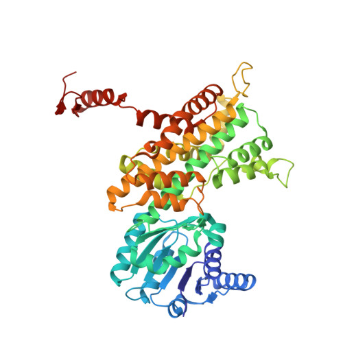
    The enzyme 6-phosphogluconate dehydrogenase (6PGD) of the malaria parasite Plasmodium falciparum catalyzes the third step of the pentose phosphate pathway converting 6-phosphogluconate (6PG) to ribulose 5-phosphate. The NADPH produced by 6PGD is crucial for antioxidant defense and redox regulation, and ribose 5-phosphate is essential for DNA and RNA synthesis in the rapidly growing parasite. Thus, 6PGD represents an attractive antimalarial drug target. In this study, we present the X-ray structures of Pf6PGD in native form as well as in complex with 6PG or nicotinamide adenine dinucleotide phosphate (NADP + ) at resolutions of 2.8, 1.9, and 2.9 Å, respectively. The overall structure of the protein is similar to structures of 6PGDs from other species; however, a flexible loop close to the active site rearranges upon binding of 6PG and likely regulates the conformation of the cofactor NADP + . Upon binding of 6PG, the active site loop adopts a closed conformation. In the absence of 6PG, the loop opens and NADP + is bound in a waiting position, indicating that the cofactor and 6PG bind independently from each other. This sequential binding mechanism was supported by kinetic studies on the homodimeric wild-type Pf6PGD. Furthermore, the function of the Plasmodium-specific residue W104L mutant was characterized by site-directed mutagenesis. Notably, the activity of Pf6PGD was found to be post-translationally redox regulated via S-nitrosylation, and screening the Medicines for Malaria Venture Malaria Box identified several compounds with IC 50 s in the low micromolar range. Together with the three-dimensional structure of the protein, this is a promising starting point for further drug discovery approaches.


  • Organizational Affiliation
    • Biochemistry and Molecular Biology, Interdisciplinary Research Center, Justus Liebig University, 35392 Giessen, Germany.

Macromolecule Content 

  • Total Structure Weight: 108.99 kDa 
  • Atom Count: 7,920 
  • Modeled Residue Count: 936 
  • Deposited Residue Count: 936 
  • Unique protein chains: 1

Macromolecules

Find similar proteins by:|  3D Structure
Entity ID: 1
MoleculeChains  Sequence LengthOrganismDetailsImage
6-phosphogluconate dehydrogenase, decarboxylating
A, B
468Plasmodium falciparum 3D7Mutation(s): 0 
Gene Names: PF14_0520PF3D7_1454700
EC: 1.1.1.44
UniProt
Find proteins for Q8IKT2 (Plasmodium falciparum (isolate 3D7))
Explore Q8IKT2 
Go to UniProtKB:  Q8IKT2
Entity Groups
Sequence Clusters30% Identity50% Identity70% Identity90% Identity95% Identity100% Identity
UniProt GroupQ8IKT2
Sequence Annotations
Expand
Reference Sequence

Small Molecules

Ligands 4 Unique
IDChains Name / Formula / InChI Key2D Diagram3D Interactions
6PG
(Subject of Investigation/LOI)

Query on 6PG


Download:Ideal Coordinates CCD File
C [auth A],
P [auth B]
6-PHOSPHOGLUCONIC ACID
C6 H13 O10 P
BIRSGZKFKXLSJQ-SQOUGZDYSA-N
PGE

Query on PGE


Download:Ideal Coordinates CCD File
Z [auth B]TRIETHYLENE GLYCOL
C6 H14 O4
ZIBGPFATKBEMQZ-UHFFFAOYSA-N
PEG

Query on PEG


Download:Ideal Coordinates CCD File
AA [auth B],
BA [auth B],
N [auth A],
O [auth A]
DI(HYDROXYETHYL)ETHER
C4 H10 O3
MTHSVFCYNBDYFN-UHFFFAOYSA-N
GOL

Query on GOL


Download:Ideal Coordinates CCD File
D [auth A]
E [auth A]
F [auth A]
G [auth A]
H [auth A]
D [auth A],
E [auth A],
F [auth A],
G [auth A],
H [auth A],
I [auth A],
J [auth A],
K [auth A],
L [auth A],
M [auth A],
Q [auth B],
R [auth B],
S [auth B],
T [auth B],
U [auth B],
V [auth B],
W [auth B],
X [auth B],
Y [auth B]
GLYCEROL
C3 H8 O3
PEDCQBHIVMGVHV-UHFFFAOYSA-N

Experimental Data & Validation

Experimental Data

  • Method: X-RAY DIFFRACTION
  • Resolution: 1.90 Å
  • R-Value Free:  0.229 (Depositor), 0.235 (DCC) 
  • R-Value Work:  0.198 (Depositor), 0.202 (DCC) 
  • R-Value Observed: 0.198 (Depositor) 
Space Group: P 32 2 1
Unit Cell:
Length ( Å )Angle ( ˚ )
a = 99.66α = 90
b = 99.66β = 90
c = 180.78γ = 120
Software Package:
Software NamePurpose
PHENIXrefinement
XDSdata reduction
XSCALEdata scaling
PHENIXphasing

Structure Validation

View Full Validation Report


Ligand Structure Quality Assessment 


Entry History 

& Funding Information

Deposition Data


Funding OrganizationLocationGrant Number
German Research FoundationGermanyBE1540/23-2
Hessian Excellence ProgrammGermanyLoewe center Druid, B3 and E3

Revision History  (Full details and data files)

  • Version 1.0: 2018-08-22
    Type: Initial release
  • Version 1.1: 2018-10-24
    Changes: Data collection, Database references
  • Version 1.2: 2024-01-17
    Changes: Data collection, Database references, Refinement description