


Phosphotriesterase PTE_A53_4
Dym, O., Aggarwal, N., Albeck, S., Unger, T., Hamer Rogotner, S., Silman, I., Leader, H., Ashani, Y., Goldsmith, M., Greisen, P., Tawfik, D., Sussman, L.J.To be published.
Experimental Data Snapshot
Starting Model: experimental
View more details
Entity ID: 1 | |||||
|---|---|---|---|---|---|
| Molecule | Chains | Sequence Length | Organism | Details | Image |
| Parathion hydrolase | 340 | Brevundimonas diminuta | Mutation(s): 0 Gene Names: opd EC: 3.1.8.1 | 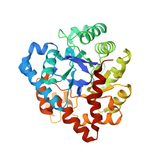 | |
UniProt | |||||
Entity Groups | |||||
| Sequence Clusters | 30% Identity50% Identity70% Identity90% Identity95% Identity100% Identity | ||||
| UniProt Group | P0A434 | ||||
Sequence AnnotationsExpand | |||||
Reference Sequence | |||||
Entity ID: 2 | |||||
|---|---|---|---|---|---|
| Molecule | Chains | Sequence Length | Organism | Details | Image |
| Parathion hydrolase | 340 | Brevundimonas diminuta | Mutation(s): 0 Gene Names: opd EC: 3.1.8.1 |  | |
UniProt | |||||
Entity Groups | |||||
| Sequence Clusters | 30% Identity50% Identity70% Identity90% Identity95% Identity100% Identity | ||||
| UniProt Group | P0A434 | ||||
Sequence AnnotationsExpand | |||||
Reference Sequence | |||||
| Ligands 4 Unique | |||||
|---|---|---|---|---|---|
| ID | Chains | Name / Formula / InChI Key | 2D Diagram | 3D Interactions | |
| D6K (Subject of Investigation/LOI) Download:Ideal Coordinates CCD File | F [auth A], K [auth B] | (4~{S},6~{R})-2,2,6-trimethyl-1,3-dioxan-4-ol C7 H14 O3 YZGBOJCVSXYSKT-RITPCOANSA-N |  | ||
| PEG (Subject of Investigation/LOI) Download:Ideal Coordinates CCD File | G [auth A] | DI(HYDROXYETHYL)ETHER C4 H10 O3 MTHSVFCYNBDYFN-UHFFFAOYSA-N |  | ||
| ZN Download:Ideal Coordinates CCD File | D [auth A], E [auth A], I [auth B], J [auth B] | ZINC ION Zn PTFCDOFLOPIGGS-UHFFFAOYSA-N |  | ||
| FMT Download:Ideal Coordinates CCD File | C [auth A], H [auth B] | FORMIC ACID C H2 O2 BDAGIHXWWSANSR-UHFFFAOYSA-N |  | ||
| Length ( Å ) | Angle ( ˚ ) |
|---|---|
| a = 54.55 | α = 90 |
| b = 81.3 | β = 94.96 |
| c = 70.61 | γ = 90 |
| Software Name | Purpose |
|---|---|
| PHENIX | refinement |
| HKL-3000 | data reduction |
| SCALEPACK | data scaling |
| PHASER | phasing |
| Funding Organization | Location | Grant Number |
|---|---|---|
| DTRA | United States | HDTRA1-11-C-0026). |
| DTRA | United States | CB10265 / HDTRA1724528 |


