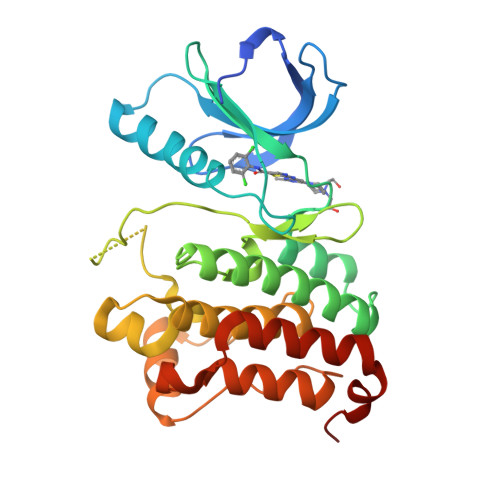
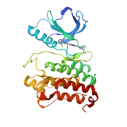
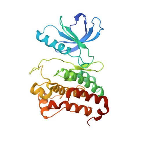
6FNM

Crystal Structure of Ephrin B4 (EphB4) Receptor Protein Kinase with Dasatinib


Experimental Data Snapshot

  • Method: X-RAY DIFFRACTION
  • Resolution: 1.16 Å
  • R-Value Free: 0.195 
  • R-Value Work: 0.184 
  • R-Value Observed: 0.184 

Starting Model: experimental
View more details

wwPDB Validation   3D Report Full Report


Ligand Structure Quality Assessment 


This is version 1.2 of the entry. See complete history


Literature

NVP-BHG712: Effects of Regioisomers on the Affinity and Selectivity toward the EPHrin Family.

Troster, A.Heinzlmeir, S.Berger, B.T.Gande, S.L.Saxena, K.Sreeramulu, S.Linhard, V.Nasiri, A.H.Bolte, M.Muller, S.Kuster, B.Medard, G.Kudlinzki, D.Schwalbe, H.

(2018) ChemMedChem 13: 1629-1633

  • DOI: https://doi.org/10.1002/cmdc.201800398
  • Primary Citation of Related Structures:  
    6FNF, 6FNG, 6FNH, 6FNI, 6FNJ, 6FNK, 6FNL, 6FNM

  • PubMed Abstract: 

    Erythropoietin-producing hepatocellular (EPH) receptors are transmembrane receptor tyrosine kinases. Their extracellular domains bind specifically to ephrin A/B ligands, and this binding modulates intracellular kinase activity. EPHs are key players in bidirectional intercellular signaling, controlling cell morphology, adhesion, and migration. They are increasingly recognized as cancer drug targets. We analyzed the binding of NVP-BHG712 (NVP) to EPHA2 and EPHB4. Unexpectedly, all tested commercially available NVP samples turned out to be a regioisomer (NVPiso) of the inhibitor, initially described in a Novartis patent application. They only differ by the localization of a single methyl group on either one of two adjacent nitrogen atoms. The two compounds of identical mass revealed different binding modes. Furthermore, both in vitro and in vivo experiments showed that the isomers differ in their kinase affinity and selectivity.


  • Organizational Affiliation

    Center for Biomolecular Magnetic Resonance (BMRZ), Institute for Organic Chemistry and Chemical Biology, Johann Wolfgang Goethe University, Max-von-Laue-Straße 7, 60438, Frankfurt am Main, Germany.


Macromolecules
Find similar proteins by:  (by identity cutoff)  |  3D Structure
Entity ID: 1
MoleculeChains Sequence LengthOrganismDetailsImage
Ephrin type-B receptor 4297Homo sapiensMutation(s): 3 
Gene Names: EPHB4HTKMYK1TYRO11
EC: 2.7.10.1
UniProt & NIH Common Fund Data Resources
Find proteins for P54760 (Homo sapiens)
Explore P54760 
Go to UniProtKB:  P54760
PHAROS:  P54760
GTEx:  ENSG00000196411 
Entity Groups  
Sequence Clusters30% Identity50% Identity70% Identity90% Identity95% Identity100% Identity
UniProt GroupP54760
Sequence Annotations
Expand
  • Reference Sequence
Small Molecules
Ligands 1 Unique
IDChains Name / Formula / InChI Key2D Diagram3D Interactions
1N1
Query on 1N1

Download Ideal Coordinates CCD File 
B [auth A]N-(2-CHLORO-6-METHYLPHENYL)-2-({6-[4-(2-HYDROXYETHYL)PIPERAZIN-1-YL]-2-METHYLPYRIMIDIN-4-YL}AMINO)-1,3-THIAZOLE-5-CARBOXAMIDE
C22 H26 Cl N7 O2 S
ZBNZXTGUTAYRHI-UHFFFAOYSA-N
Experimental Data & Validation

Experimental Data

  • Method: X-RAY DIFFRACTION
  • Resolution: 1.16 Å
  • R-Value Free: 0.195 
  • R-Value Work: 0.184 
  • R-Value Observed: 0.184 
  • Space Group: P 1 21 1
Unit Cell:
Length ( Å )Angle ( ˚ )
a = 45.941α = 90
b = 54.111β = 110.45
c = 62.162γ = 90
Software Package:
Software NamePurpose
PHENIXrefinement
XDSdata reduction
Aimlessdata scaling
PHASERphasing

Structure Validation

View Full Validation Report


Ligand Structure Quality Assessment 


Entry History & Funding Information

Deposition Data


Funding OrganizationLocationGrant Number
DKTKGermanyL590

Revision History  (Full details and data files)

  • Version 1.0: 2018-08-08
    Type: Initial release
  • Version 1.1: 2018-08-29
    Changes: Data collection, Database references
  • Version 1.2: 2024-01-17
    Changes: Data collection, Database references, Refinement description