6FNA | pdb_00006fna

Mono- and bivalent 14-3-3 inhibitors for characterizing supramolecular lysine-PEG interactions in proteins


Experimental Data Snapshot

  • Method: X-RAY DIFFRACTION
  • Resolution: 2.12 Å
  • R-Value Free: 
    0.236 (Depositor), 0.238 (DCC) 
  • R-Value Work: 
    0.202 (Depositor), 0.208 (DCC) 
  • R-Value Observed: 
    0.203 (Depositor) 

Starting Model: experimental
View more details

wwPDB Validation 3D Report Full Report

Validation slider image for 6FNA

Ligand Structure Quality Assessment 


This is version 1.3 of the entry. See complete history

Literature

Mono- and Bivalent 14-3-3 Inhibitors for Characterizing Supramolecular "Lysine Wrapping" of Oligoethylene Glycol (OEG) Moieties in Proteins.

Yilmaz, E.Bier, D.Guillory, X.Briels, J.Ruiz-Blanco, Y.B.Sanchez-Garcia, E.Ottmann, C.Kaiser, M.

(2018) Chemistry 24: 13807-13814

  • DOI: https://doi.org/10.1002/chem.201801074
  • Primary Citation Related Structures: 
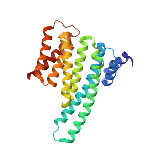
    6FN9, 6FNA, 6FNB, 6FNC

  • PubMed Abstract: 

    Previous studies have indicated the presence of defined interactions between oligo or poly(ethylene glycol) (OEG or PEG) and lysine residues. In these interactions, the OEG or PEG residues "wrap around" the lysine amino group, thereby enabling complexation of the amino group by the ether oxygen residues. The resulting biochemical binding affinity and thus biological relevance of this supramolecular interaction however remains unclear so far. Here, we report that OEG-containing phosphophenol ether inhibitors of 14-3-3 proteins also display such a "lysine-wrapping" binding mode. For better investigating the biochemical relevance of this binding mode, we made use of the dimeric nature of 14-3-3 proteins and designed as well as synthesized a set of bivalent 14-3-3 inhibitors for biochemical and X-ray crystallography-based structural studies. We found that all synthesized derivatives adapted the "lysine-wrapping" binding mode in the crystal structures; in solution, a different binding mode is however observed, most probably as the "lysine-wrapping" binding mode turned out to be a rather weak interaction. Accordingly, our studies demonstrate that structural studies of OEG-lysine interactions are difficult to interpret and their presence in structural studies may not automatically be correlated with a relevant interaction also in solution but requires further biochemical studies.


  • Organizational Affiliation
    • Chemical Biology, Zentrum für Medizinische Biotechnologie, Fakultät für Biologie, Universität Duisburg-Essen, Universitätsstr. 2, 45117, Essen, Germany.

Macromolecule Content 

  • Total Structure Weight: 55.55 kDa 
  • Atom Count: 4,054 
  • Modeled Residue Count: 457 
  • Deposited Residue Count: 460 
  • Unique protein chains: 1

Macromolecules

Find similar proteins by:|  3D Structure
Entity ID: 1
MoleculeChains  Sequence LengthOrganismDetailsImage
14-3-3 protein zeta/delta
A, B
230Homo sapiensMutation(s): 0 
Gene Names: YWHAZ
UniProt & NIH Common Fund Data Resources
Find proteins for P63104 (Homo sapiens)
Explore P63104 
Go to UniProtKB:  P63104
PHAROS:  P63104
GTEx:  ENSG00000164924 
Entity Groups
Sequence Clusters30% Identity50% Identity70% Identity90% Identity95% Identity100% Identity
UniProt GroupP63104
Sequence Annotations
Expand
Reference Sequence

Small Molecules

Ligands 5 Unique
IDChains Name / Formula / InChI Key2D Diagram3D Interactions
DWE

Query on DWE


Download:Ideal Coordinates CCD File
C [auth A],
J [auth B]
[2-[2-oxidanylidene-2-[[3-[2-[2-[2-[3-oxidanylidene-3-[2-[3-[2-[2-[2-[[4-[2-(2-phosphonophenoxy)ethanoylamino]phenyl]carbonylamino]ethoxy]ethoxy]ethoxy]propanoylamino]ethylamino]propoxy]ethoxy]ethoxy]ethylcarbamoyl]phenyl]amino]ethoxy]phenyl]phosphonic acid
C50 H66 N6 O20 P2
IJJNCSHPFORWPU-UHFFFAOYSA-N
BEZ

Query on BEZ


Download:Ideal Coordinates CCD File
D [auth A],
K [auth B]
BENZOIC ACID
C7 H6 O2
WPYMKLBDIGXBTP-UHFFFAOYSA-N
GOL

Query on GOL


Download:Ideal Coordinates CCD File
H [auth A],
I [auth A]
GLYCEROL
C3 H8 O3
PEDCQBHIVMGVHV-UHFFFAOYSA-N
CA

Query on CA


Download:Ideal Coordinates CCD File
E [auth A],
L [auth B]
CALCIUM ION
Ca
BHPQYMZQTOCNFJ-UHFFFAOYSA-N
CL

Query on CL


Download:Ideal Coordinates CCD File
F [auth A],
G [auth A],
M [auth B],
N [auth B]
CHLORIDE ION
Cl
VEXZGXHMUGYJMC-UHFFFAOYSA-M

Experimental Data & Validation

Experimental Data

  • Method: X-RAY DIFFRACTION
  • Resolution: 2.12 Å
  • R-Value Free:  0.236 (Depositor), 0.238 (DCC) 
  • R-Value Work:  0.202 (Depositor), 0.208 (DCC) 
  • R-Value Observed: 0.203 (Depositor) 
Space Group: P 21 21 21
Unit Cell:
Length ( Å )Angle ( ˚ )
a = 72.035α = 90
b = 102.888β = 90
c = 113.627γ = 90
Software Package:
Software NamePurpose
XDSdata reduction
XSCALEdata scaling
PHASERphasing
REFMACrefinement
PDB_EXTRACTdata extraction

Structure Validation

View Full Validation Report


Ligand Structure Quality Assessment 


Entry History 

& Funding Information

Deposition Data


Funding OrganizationLocationGrant Number
German Research FoundationGermanyCRC1093 (Supramolecular Chemistry on Proteins)

Revision History  (Full details and data files)

  • Version 1.0: 2018-07-04
    Type: Initial release
  • Version 1.1: 2018-10-03
    Changes: Data collection, Database references
  • Version 1.2: 2018-12-19
    Changes: Data collection, Derived calculations
  • Version 1.3: 2024-01-17
    Changes: Data collection, Database references, Refinement description