Download Mendeley
De novoprotein structure determination by heavy-atom soaking in lipidic cubic phase and SIRAS phasing using serial synchrotron crystallography.
Botha, S., Baitan, D., Jungnickel, K.E.J., Oberthur, D., Schmidt, C., Stern, S., Wiedorn, M.O., Perbandt, M., Chapman, H.N., Betzel, C.(2018) IUCrJ 5: 524-530
- PubMed: 30224955 Search on PubMedSearch on PubMed Central
- DOI: https://doi.org/10.1107/S2052252518009223
- Primary Citation Related Structures:
6FJS - PubMed Abstract:
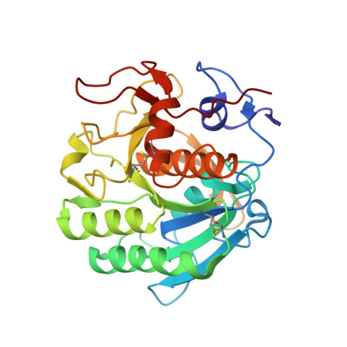
During the past few years, serial crystallography methods have undergone continuous development and serial data collection has become well established at high-intensity synchrotron-radiation beamlines and XFEL radiation sources. However, the application of experimental phasing to serial crystallography data has remained a challenging task owing to the inherent inaccuracy of the diffraction data. Here, a particularly gentle method for incorporating heavy atoms into micrometre-sized crystals utilizing lipidic cubic phase (LCP) as a carrier medium is reported. Soaking in LCP prior to data collection offers a new, efficient and gentle approach for preparing heavy-atom-derivative crystals directly before diffraction data collection using serial crystallography methods. This approach supports effective phasing by utilizing a reasonably low number of diffraction patterns. Using synchrotron radiation and exploiting the anomalous scattering signal of mercury for single isomorphous replacement with anomalous scattering (SIRAS) phasing resulted in high-quality electron-density maps that were sufficient for building a complete structural model of proteinase K at 1.9 Å resolution using automatic model-building tools.
- Institute of Biochemistry and Molecular Biology, Chemistry Department, University of Hamburg, Martin-Luther-King Platz 6, 20146 Hamburg, Germany.
Organizational Affiliation: