6FJO | pdb_00006fjo

Adenovirus species 26 knob protein, very high resolution


Experimental Data Snapshot

  • Method: X-RAY DIFFRACTION
  • Resolution: 1.17 Å
  • R-Value Free: 
    0.190 (Depositor), 0.193 (DCC) 
  • R-Value Work: 
    0.169 (Depositor), 0.175 (DCC) 
  • R-Value Observed: 
    0.170 (Depositor) 

Starting Model: experimental
View more details

wwPDB Validation 3D Report Full Report

Validation slider image for 6FJO

This is version 1.5 of the entry. See complete history

Literature

Human adenovirus type 26 uses sialic acid-bearing glycans as a primary cell entry receptor.

Baker, A.T.Mundy, R.M.Davies, J.A.Rizkallah, P.J.Parker, A.L.

(2019) Sci Adv 5: eaax3567-eaax3567

  • DOI: https://doi.org/10.1126/sciadv.aax3567
  • Primary Citation Related Structures: 
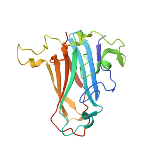
    6FJO, 6QU6, 6QU8

  • PubMed Abstract: 

    Adenoviruses are clinically important agents. They cause respiratory distress, gastroenteritis, and epidemic keratoconjunctivitis. As non-enveloped, double-stranded DNA viruses, they are easily manipulated, making them popular vectors for therapeutic applications, including vaccines. Species D adenovirus type 26 (HAdV-D26) is both a cause of EKC and other diseases and a promising vaccine vector. HAdV-D26-derived vaccines are under investigation as protective platforms against HIV, Zika, and respiratory syncytial virus infections and are in phase 3 clinical trials for Ebola. We recently demonstrated that HAdV-D26 does not use CD46 or Desmoglein-2 as entry receptors, while the putative interaction with coxsackie and adenovirus receptor is low affinity and unlikely to represent the primary cell receptor. Here, we establish sialic acid as a primary entry receptor used by HAdV-D26. We demonstrate that removal of cell surface sialic acid inhibits HAdV-D26 infection, and provide a high-resolution crystal structure of HAdV-D26 fiber-knob in complex with sialic acid.


  • Organizational Affiliation
    • Division of Cancer and Genetics, School of Medicine, Cardiff University, Heath Park, Cardiff CF14 4XN, UK.

Macromolecule Content 

  • Total Structure Weight: 20.98 kDa 
  • Atom Count: 1,761 
  • Modeled Residue Count: 183 
  • Deposited Residue Count: 183 
  • Unique protein chains: 1

Macromolecules

Find similar proteins by:|  3D Structure
Entity ID: 1
MoleculeChains  Sequence LengthOrganismDetailsImage
Fiber183Human adenovirus 26Mutation(s): 0 
UniProt
Find proteins for A4ZKM1 (Human adenovirus 26)
Explore A4ZKM1 
Go to UniProtKB:  A4ZKM1
Entity Groups
Sequence Clusters30% Identity50% Identity70% Identity90% Identity95% Identity100% Identity
UniProt GroupA4ZKM1
Sequence Annotations
Expand
Reference Sequence

Experimental Data & Validation

Experimental Data

  • Method: X-RAY DIFFRACTION
  • Resolution: 1.17 Å
  • R-Value Free:  0.190 (Depositor), 0.193 (DCC) 
  • R-Value Work:  0.169 (Depositor), 0.175 (DCC) 
  • R-Value Observed: 0.170 (Depositor) 
Space Group: P 21 3
Unit Cell:
Length ( Å )Angle ( ˚ )
a = 85.776α = 90
b = 85.776β = 90
c = 85.776γ = 90
Software Package:
Software NamePurpose
REFMACrefinement
xia2data reduction
XDSdata reduction
Aimlessdata scaling
PHASERphasing

Structure Validation

View Full Validation Report


Entry History 

Deposition Data

Revision History  (Full details and data files)

  • Version 1.0: 2019-02-06
    Type: Initial release
  • Version 1.1: 2019-02-13
    Changes: Data collection, Database references
  • Version 1.2: 2019-05-01
    Changes: Data collection, Database references
  • Version 1.3: 2019-09-18
    Changes: Data collection, Database references
  • Version 1.4: 2019-09-25
    Changes: Data collection, Database references
  • Version 1.5: 2024-01-17
    Changes: Data collection, Database references, Refinement description