6FEK | pdb_00006fek

Oncogenic point mutation of RET receptor tyrosine kinase


Experimental Data Snapshot

  • Method: X-RAY DIFFRACTION
  • Resolution: 2.30 Å
  • R-Value Free: 
    0.244 (Depositor), 0.244 (DCC) 
  • R-Value Work: 
    0.201 (Depositor), 0.205 (DCC) 
  • R-Value Observed: 
    0.203 (Depositor) 

wwPDB Validation 3D Report Full Report

Validation slider image for 6FEK

Ligand Structure Quality Assessment 


This is version 1.2 of the entry. See complete history

Literature

A secondary RET mutation in the activation loop conferring resistance to vandetanib.

Nakaoku, T.Kohno, T.Araki, M.Niho, S.Chauhan, R.Knowles, P.P.Tsuchihara, K.Matsumoto, S.Shimada, Y.Mimaki, S.Ishii, G.Ichikawa, H.Nagatoishi, S.Tsumoto, K.Okuno, Y.Yoh, K.McDonald, N.Q.Goto, K.

(2018) Nat Commun 9: 625-625

  • DOI: https://doi.org/10.1038/s41467-018-02994-7
  • Primary Citation Related Structures: 
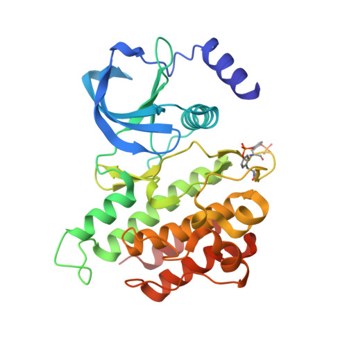
    6FEK

  • PubMed Abstract: 

    Resistance to vandetanib, a type I RET kinase inhibitor, developed in a patient with metastatic lung adenocarcinoma harboring a CCDC6-RET fusion that initially exhibited a response to treatment. The resistant tumor acquired a secondary mutation resulting in a serine-to-phenylalanine substitution at codon 904 in the activation loop of the RET kinase domain. The S904F mutation confers resistance to vandetanib by increasing the ATP affinity and autophosphorylation activity of RET kinase. A reduced interaction with the drug is also observed in vitro for the S904F mutant by thermal shift assay. A crystal structure of the S904F mutant reveals a small hydrophobic core around F904 likely to enhance basal kinase activity by stabilizing an active conformer. Our findings indicate that missense mutations in the activation loop of the kinase domain are able to increase kinase activity and confer drug resistance through allosteric effects.


  • Organizational Affiliation
    • Division of Genome Biology, National Cancer Center Research Institute, 5-1-1, Tsukiji, Chuo-ku, Tokyo, 1040045, Japan.

Macromolecule Content 

  • Total Structure Weight: 34.71 kDa 
  • Atom Count: 2,343 
  • Modeled Residue Count: 296 
  • Deposited Residue Count: 299 
  • Unique protein chains: 1

Macromolecules

Find similar proteins by:|  3D Structure
Entity ID: 1
MoleculeChains  Sequence LengthOrganismDetailsImage
Proto-oncogene tyrosine-protein kinase receptor Ret299Homo sapiensMutation(s): 1 
Gene Names: RETCDHF12CDHR16PTCRET51
EC: 2.7.10.1
UniProt & NIH Common Fund Data Resources
Find proteins for P07949 (Homo sapiens)
Explore P07949 
Go to UniProtKB:  P07949
PHAROS:  P07949
GTEx:  ENSG00000165731 
Entity Groups
Sequence Clusters30% Identity50% Identity70% Identity90% Identity95% Identity100% Identity
UniProt GroupP07949
Sequence Annotations
Expand
Reference Sequence

Experimental Data & Validation

Experimental Data

  • Method: X-RAY DIFFRACTION
  • Resolution: 2.30 Å
  • R-Value Free:  0.244 (Depositor), 0.244 (DCC) 
  • R-Value Work:  0.201 (Depositor), 0.205 (DCC) 
  • R-Value Observed: 0.203 (Depositor) 
Space Group: P 43 21 2
Unit Cell:
Length ( Å )Angle ( ˚ )
a = 50.901α = 90
b = 50.901β = 90
c = 242.515γ = 90
Software Package:
Software NamePurpose
PHENIXrefinement
XDSdata reduction
XDSdata scaling
PHENIXphasing

Structure Validation

View Full Validation Report


Ligand Structure Quality Assessment 


Entry History 

& Funding Information

Deposition Data


Funding OrganizationLocationGrant Number
Francis Crick InstituteUnited KingdomFC001115

Revision History  (Full details and data files)

  • Version 1.0: 2018-02-28
    Type: Initial release
  • Version 1.1: 2018-05-02
    Changes: Data collection
  • Version 1.2: 2024-10-16
    Changes: Advisory, Data collection, Database references, Structure summary