6FB3 | pdb_00006fb3

Teneurin 2 Partial Extracellular Domain


Experimental Data Snapshot

  • Method: X-RAY DIFFRACTION
  • Resolution: 2.38 Å
  • R-Value Free: 
    0.242 (Depositor), 0.262 (DCC) 
  • R-Value Work: 
    0.225 (Depositor), 0.245 (DCC) 
  • R-Value Observed: 
    0.226 (Depositor) 

wwPDB Validation 3D Report Full Report

Validation slider image for 6FB3

Ligand Structure Quality Assessment 


This is version 2.1 of the entry. See complete history

Literature

Structures of Teneurin adhesion receptors reveal an ancient fold for cell-cell interaction.

Jackson, V.A.Meijer, D.H.Carrasquero, M.van Bezouwen, L.S.Lowe, E.D.Kleanthous, C.Janssen, B.J.C.Seiradake, E.

(2018) Nat Commun 9: 1079-1079

  • DOI: https://doi.org/10.1038/s41467-018-03460-0
  • Primary Citation Related Structures: 
    6FAY, 6FB3

  • PubMed Abstract: 

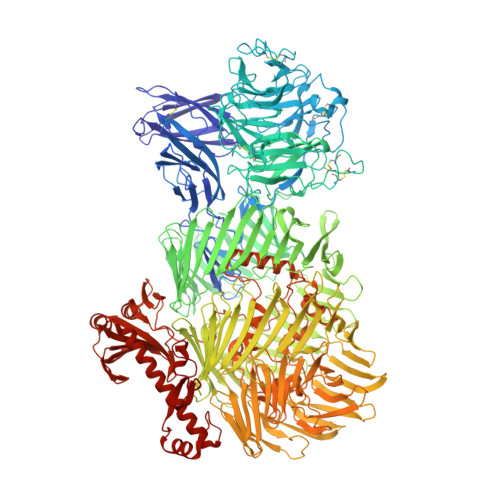
    Teneurins are ancient cell-cell adhesion receptors that are vital for brain development and synapse organisation. They originated in early metazoan evolution through a horizontal gene transfer event when a bacterial YD-repeat toxin fused to a eukaryotic receptor. We present X-ray crystallography and cryo-EM structures of two Teneurins, revealing a ~200 kDa extracellular super-fold in which eight sub-domains form an intricate structure centred on a spiralling YD-repeat shell. An alternatively spliced loop, which is implicated in homophilic Teneurin interaction and specificity, is exposed and thus poised for interaction. The N-terminal side of the shell is 'plugged' via a fibronectin-plug domain combination, which defines a new class of YD proteins. Unexpectedly, we find that these proteins are widespread amongst modern bacteria, suggesting early metazoan receptor evolution from a distinct class of proteins, which today includes both bacterial proteins and eukaryotic Teneurins.


  • Organizational Affiliation
    • Department of Biochemistry, Oxford University, OX1 3QU, Oxford, UK. verity.jackson@magd.ox.ac.uk.

Macromolecule Content 

  • Total Structure Weight: 853.68 kDa 
  • Atom Count: 59,451 
  • Modeled Residue Count: 7,344 
  • Deposited Residue Count: 7,436 
  • Unique protein chains: 1

Macromolecules

Find similar proteins by:|  3D Structure
Entity ID: 1
MoleculeChains  Sequence LengthOrganismDetailsImage
Teneurin-2
A, B, C, D
1,859Gallus gallusMutation(s): 0 
Gene Names: TENM2ODZ2TNM2
UniProt
Find proteins for Q9DER5 (Gallus gallus)
Explore Q9DER5 
Go to UniProtKB:  Q9DER5
Entity Groups
Sequence Clusters30% Identity50% Identity70% Identity90% Identity95% Identity100% Identity
UniProt GroupQ9DER5
Glycosylation
Glycosylation Sites: 11
Sequence Annotations
Expand
Reference Sequence

Oligosaccharides

Help  
Entity ID: 2
MoleculeChains Length2D Diagram GlycosylationD Interactions
alpha-D-mannopyranose-(1-2)-alpha-D-mannopyranose-(1-2)-alpha-D-mannopyranose-(1-3)-[alpha-D-mannopyranose-(1-3)-alpha-D-mannopyranose-(1-6)]beta-D-mannopyranose-(1-4)-2-acetamido-2-deoxy-beta-D-glucopyranose-(1-4)-2-acetamido-2-deoxy-beta-D-glucopyranose
E, K, Q, W
8N-Glycosylation
Glycosylation Resources
GlyTouCan: G83582BK
GlyCosmos: G83582BK
GlyGen: G83582BK

Small Molecules

Ligands 1 Unique
IDChains Name / Formula / InChI Key2D Diagram3D Interactions
NAG

Query on NAG


Download:Ideal Coordinates CCD File
CA [auth A]
DA [auth A]
EA [auth A]
FA [auth A]
GA [auth A]
CA [auth A],
DA [auth A],
EA [auth A],
FA [auth A],
GA [auth A],
HA [auth B],
IA [auth B],
JA [auth B],
KA [auth B],
LA [auth B],
MA [auth C],
NA [auth C],
OA [auth C],
PA [auth C],
QA [auth C],
RA [auth D],
SA [auth D],
TA [auth D],
UA [auth D],
VA [auth D]
2-acetamido-2-deoxy-beta-D-glucopyranose
C8 H15 N O6
OVRNDRQMDRJTHS-FMDGEEDCSA-N

Experimental Data & Validation

Experimental Data

  • Method: X-RAY DIFFRACTION
  • Resolution: 2.38 Å
  • R-Value Free:  0.242 (Depositor), 0.262 (DCC) 
  • R-Value Work:  0.225 (Depositor), 0.245 (DCC) 
  • R-Value Observed: 0.226 (Depositor) 
Space Group: P 1 21 1
Unit Cell:
Length ( Å )Angle ( ˚ )
a = 88.56α = 90
b = 452.56β = 95.12
c = 146.36γ = 90
Software Package:
Software NamePurpose
BUSTERrefinement
XDSdata reduction
XDSdata scaling
PHASERphasing

Structure Validation

View Full Validation Report


Ligand Structure Quality Assessment 


Entry History 

& Funding Information

Deposition Data


Funding OrganizationLocationGrant Number
Wellcome TrustUnited Kingdom202827/Z/16/Z
Medical Research Council (United Kingdom)United KingdomMR/L018039/1

Revision History  (Full details and data files)

  • Version 1.0: 2018-03-28
    Type: Initial release
  • Version 1.1: 2019-03-27
    Changes: Data collection, Derived calculations, Other
  • Version 1.2: 2019-07-10
    Changes: Data collection
  • Version 2.0: 2020-07-29
    Type: Remediation
    Reason: Carbohydrate remediation
    Changes: Atomic model, Data collection, Derived calculations, Structure summary
  • Version 2.1: 2024-11-06
    Changes: Data collection, Database references, Derived calculations, Structure summary