6FAH | pdb_00006fah

Molecular basis of the flavin-based electron-bifurcating caffeyl-CoA reductase reaction


Experimental Data Snapshot

  • Method: X-RAY DIFFRACTION
  • Resolution: 3.13 Å
  • R-Value Free: 
    0.286 (Depositor), 0.293 (DCC) 
  • R-Value Work: 
    0.234 (Depositor), 0.240 (DCC) 
  • R-Value Observed: 
    0.237 (Depositor) 

Starting Models: experimental
View more details

wwPDB Validation 3D Report Full Report

Validation slider image for 6FAH

Ligand Structure Quality Assessment 


This is version 1.3 of the entry. See complete history

Literature

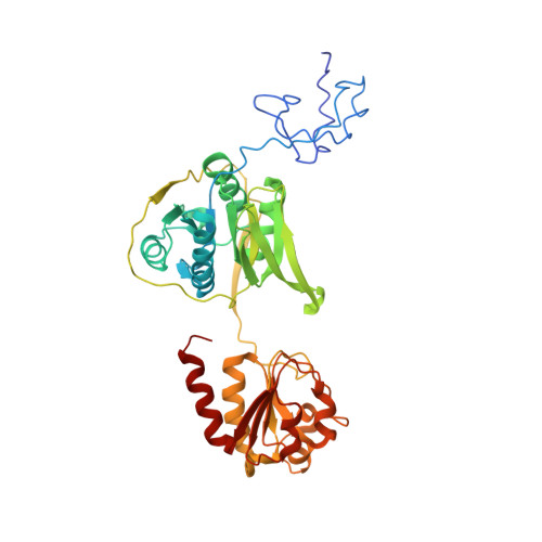
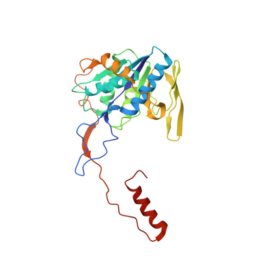
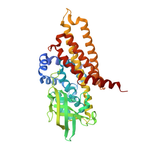
Molecular basis of the flavin-based electron-bifurcating caffeyl-CoA reductase reaction.

Demmer, J.K.Bertsch, J.Oppinger, C.Wohlers, H.Kayastha, K.Demmer, U.Ermler, U.Muller, V.

(2018) FEBS Lett 592: 332-342

  • DOI: https://doi.org/10.1002/1873-3468.12971
  • Primary Citation Related Structures: 
    6FAH

  • PubMed Abstract: 

    Flavin-based electron bifurcation (FBEB) is a recently discovered mode of energy coupling in anaerobic microorganisms. The electron-bifurcating caffeyl-CoA reductase (CarCDE) catalyzes the reduction of caffeyl-CoA and ferredoxin by oxidizing NADH. The 3.5 Å structure of the heterododecameric Car(CDE) 4 complex of Acetobacterium woodii, presented here, reveals compared to other electron-transferring flavoprotein/acyl dehydrogenase family members an additional ferredoxin-like domain with two [4Fe-4S] clusters N-terminally fused to CarE. It might serve, in vivo, as specific adaptor for the physiological electron acceptor. Kinetic analysis of a CarCDE(∆Fd) complex indicates the bypassing of the ferredoxin-like domain by artificial electron acceptors. Site-directed mutagenesis studies substantiated the crucial role of the C-terminal arm of CarD and of ArgE203, hydrogen-bonded to the bifurcating FAD, for FBEB.


  • Organizational Affiliation
    • Max-Planck-Institut für Biophysik, Frankfurt am Main, Germany.

Macromolecule Content 

  • Total Structure Weight: 231.36 kDa 
  • Atom Count: 15,985 
  • Modeled Residue Count: 2,064 
  • Deposited Residue Count: 2,074 
  • Unique protein chains: 3

Macromolecules

Find similar proteins by:|  3D Structure
Entity ID: 1
MoleculeChains  Sequence LengthOrganismDetailsImage
Caffeyl-CoA reductase-Etf complex subunit CarE
A, E
396Acetobacterium woodii DSM 1030Mutation(s): 0 
EC: 1.3.1.108
UniProt
Find proteins for H6LGM8 (Acetobacterium woodii (strain ATCC 29683 / DSM 1030 / JCM 2381 / KCTC 1655 / WB1))
Explore H6LGM8 
Go to UniProtKB:  H6LGM8
Entity Groups
Sequence Clusters30% Identity50% Identity70% Identity90% Identity95% Identity100% Identity
UniProt GroupH6LGM8
Sequence Annotations
Expand
Reference Sequence
Find similar proteins by:|  3D Structure
Entity ID: 2
MoleculeChains  Sequence LengthOrganismDetailsImage
Caffeyl-CoA reductase-Etf complex subunit CarD
B, F
262Acetobacterium woodii DSM 1030Mutation(s): 0 
EC: 1.3.1.108
UniProt
Find proteins for H6LGM7 (Acetobacterium woodii (strain ATCC 29683 / DSM 1030 / JCM 2381 / KCTC 1655 / WB1))
Explore H6LGM7 
Go to UniProtKB:  H6LGM7
Entity Groups
Sequence Clusters30% Identity50% Identity70% Identity90% Identity95% Identity100% Identity
UniProt GroupH6LGM7
Sequence Annotations
Expand
Reference Sequence
Find similar proteins by:|  3D Structure
Entity ID: 3
MoleculeChains  Sequence LengthOrganismDetailsImage
Caffeyl-CoA reductase-Etf complex subunit CarC
C, D
379Acetobacterium woodii DSM 1030Mutation(s): 0 
EC: 1.3.1.108
UniProt
Find proteins for H6LGM6 (Acetobacterium woodii (strain ATCC 29683 / DSM 1030 / JCM 2381 / KCTC 1655 / WB1))
Explore H6LGM6 
Go to UniProtKB:  H6LGM6
Entity Groups
Sequence Clusters30% Identity50% Identity70% Identity90% Identity95% Identity100% Identity
UniProt GroupH6LGM6
Sequence Annotations
Expand
Reference Sequence

Small Molecules

Ligands 3 Unique
IDChains Name / Formula / InChI Key2D Diagram3D Interactions
FAD

Query on FAD


Download:Ideal Coordinates CCD File
G [auth A]
J [auth B]
K [auth C]
Q [auth D]
T [auth E]
G [auth A],
J [auth B],
K [auth C],
Q [auth D],
T [auth E],
W [auth F]
FLAVIN-ADENINE DINUCLEOTIDE
C27 H33 N9 O15 P2
VWWQXMAJTJZDQX-UYBVJOGSSA-N
SF4

Query on SF4


Download:Ideal Coordinates CCD File
H [auth A],
I [auth A],
U [auth E],
V [auth E]
IRON/SULFUR CLUSTER
Fe4 S4
LJBDFODJNLIPKO-UHFFFAOYSA-N
SO4

Query on SO4


Download:Ideal Coordinates CCD File
L [auth C]
M [auth C]
N [auth C]
O [auth C]
P [auth C]
L [auth C],
M [auth C],
N [auth C],
O [auth C],
P [auth C],
R [auth D],
S [auth D]
SULFATE ION
O4 S
QAOWNCQODCNURD-UHFFFAOYSA-L

Experimental Data & Validation

Experimental Data

  • Method: X-RAY DIFFRACTION
  • Resolution: 3.13 Å
  • R-Value Free:  0.286 (Depositor), 0.293 (DCC) 
  • R-Value Work:  0.234 (Depositor), 0.240 (DCC) 
  • R-Value Observed: 0.237 (Depositor) 
Space Group: C 1 2 1
Unit Cell:
Length ( Å )Angle ( ˚ )
a = 213.74α = 90
b = 144.26β = 98.93
c = 102.84γ = 90
Software Package:
Software NamePurpose
PHENIXrefinement
XSCALEdata scaling
XDSdata reduction
PHASERphasing
Cootmodel building

Structure Validation

View Full Validation Report


Ligand Structure Quality Assessment 


Entry History 

& Funding Information

Deposition Data


Funding OrganizationLocationGrant Number
German Research FoundationGermany--

Revision History  (Full details and data files)

  • Version 1.0: 2018-01-24
    Type: Initial release
  • Version 1.1: 2018-02-21
    Changes: Database references
  • Version 1.2: 2024-01-17
    Changes: Data collection, Database references, Derived calculations, Refinement description
  • Version 1.3: 2025-10-01
    Changes: Advisory, Derived calculations, Structure summary