6F7N | pdb_00006f7n

NMR structure of EphA2-Sam stapled peptides (S13STshort)


Experimental Data Snapshot

  • Method: SOLUTION NMR
  • Conformers Calculated: 100 
  • Conformers Submitted: 20 
  • Selection Criteria: target function 

wwPDB Validation 3D Report Full Report

Validation slider image for 6F7N

This is version 2.1 of the entry. See complete history

Literature

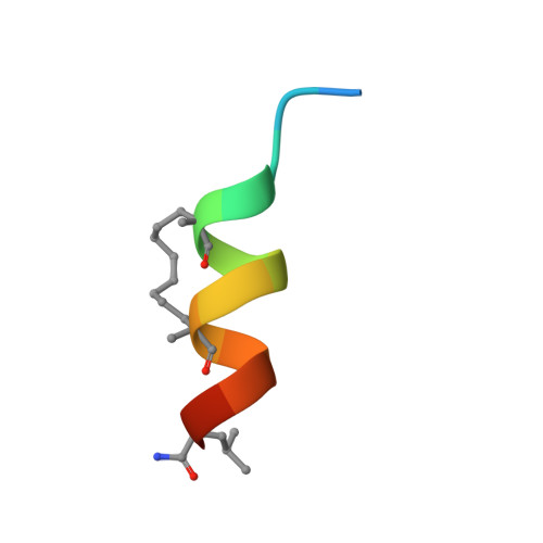
Sam domain-based stapled peptides: Structural analysis and interaction studies with the Sam domains from the EphA2 receptor and the lipid phosphatase Ship2.

Mercurio, F.A.Pirone, L.Di Natale, C.Marasco, D.Pedone, E.M.Leone, M.

(2018) Bioorg Chem 80: 602-610

  • DOI: https://doi.org/10.1016/j.bioorg.2018.07.013
  • Primary Citation Related Structures: 
    6F7M, 6F7N, 6F7O

  • PubMed Abstract: 

    Sam (Sterile alpha motif) domains represent small helical protein-protein interaction modules which play versatile functions in different cellular processes. The Sam domain from the EphA2 receptor binds the Sam domain of the lipid phosphatase Ship2 and this interaction modulates receptor endocytosis and degradation primarily generating pro-oncogenic effects in cell. To identify molecule antagonists of the EphA2-Sam/Ship2-Sam complex with anti-cancer activity, we focused on hydrocarbon helical stapled peptides. EphA2-Sam and one of its interactors (i.e., the first Sam domain of the adaptor protein Odin) were used as model systems for peptide design. Increase in helicity in the stapled peptides, with respect to the corresponding linear/native-like regions, was proved by structural studies conducted through CD (Circular Dichroism) and NMR (Nuclear Magnetic Resonance). Interestingly, interaction assays by means of NMR, SPR (Surface Plasmon Resonance) and MST (MicroScale Thermophoresis) techniques led to the discovery of a novel ligand of Ship2-Sam.


  • Organizational Affiliation
    • Institute of Biostructures and Bioimaging (CNR), Naples, Italy; InterUniversity Research Centre on Bioactive Peptides (CIRPEB), University of Naples Federico II, Naples, Italy.

Macromolecule Content 

  • Total Structure Weight: 1.58 kDa 
  • Atom Count: 113 
  • Modeled Residue Count: 15 
  • Deposited Residue Count: 15 
  • Unique protein chains: 1

Macromolecules

Find similar proteins by:  Sequence   |   3D Structure  
Entity ID: 1
MoleculeChains  Sequence LengthOrganismDetailsImage
Ephrin type-A receptor 215Homo sapiensMutation(s): 4 
EC: 2.7.10.1
UniProt & NIH Common Fund Data Resources
Find proteins for P29317 (Homo sapiens)
Explore P29317 
Go to UniProtKB:  P29317
PHAROS:  P29317
GTEx:  ENSG00000142627 
Entity Groups
Sequence Clusters30% Identity50% Identity70% Identity90% Identity95% Identity100% Identity
UniProt GroupP29317
Sequence Annotations
Expand
Reference Sequence

Small Molecules

Modified Residues  1 Unique
IDChains TypeFormula2D DiagramParent
MK8
Query on MK8
A
L-PEPTIDE LINKINGC7 H15 N O2LEU

Experimental Data & Validation

Experimental Data

  • Method: SOLUTION NMR
  • Conformers Calculated: 100 
  • Conformers Submitted: 20 
  • Selection Criteria: target function 

Structure Validation

View Full Validation Report


Entry History 

Deposition Data

Revision History  (Full details and data files)

  • Version 1.0: 2018-08-01
    Type: Initial release
  • Version 1.1: 2019-05-08
    Changes: Data collection
  • Version 1.2: 2023-06-14
    Changes: Database references, Other
  • Version 2.0: 2023-11-15
    Changes: Atomic model, Data collection
  • Version 2.1: 2024-11-20
    Changes: Database references, Structure summary