6F2P | pdb_00006f2p

Structure of Paenibacillus xanthan lyase to 2.6 A resolution


Experimental Data Snapshot

  • Method: X-RAY DIFFRACTION
  • Resolution: 2.60 Å
  • R-Value Free: 
    0.213 (Depositor), 0.216 (DCC) 
  • R-Value Work: 
    0.165 (Depositor), 0.169 (DCC) 
  • R-Value Observed: 
    0.167 (Depositor) 

Starting Model: experimental
View more details

wwPDB Validation 3D Report Full Report

Validation slider image for 6F2P

This is version 1.3 of the entry. See complete history

Literature

Structure and Dynamics of a Promiscuous Xanthan Lyase from Paenibacillus nanensis and the Design of Variants with Increased Stability and Activity.

Jensen, P.F.Kadziola, A.Comamala, G.Segura, D.R.Anderson, L.Poulsen, J.N.Rasmussen, K.K.Agarwal, S.Sainathan, R.K.Monrad, R.N.Svendsen, A.Nielsen, J.E.Lo Leggio, L.Rand, K.D.

(2019) Cell Chem Biol 26: 191-202.e6

  • DOI: https://doi.org/10.1016/j.chembiol.2018.10.016
  • Primary Citation Related Structures: 
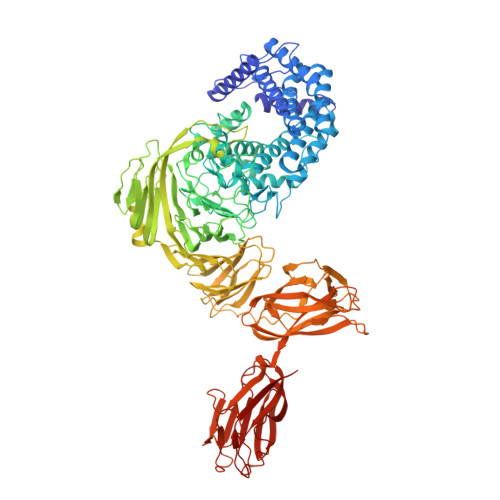
    6F2P

  • PubMed Abstract: 

    We have characterized the structure and dynamics of the carbohydrate-modifying enzyme Paenibacillus nanensis xanthan lyase (PXL) involved in the degradation of xanthan by X-ray crystallography, small-angle X-ray scattering, and hydrogen/deuterium exchange mass spectrometry. Unlike other xanthan lyases, PXL is specific for both unmodified mannose and pyruvylated mannose, which we find is correlated with structural differences in the substrate binding groove. The structure of the full-length enzyme reveals two additional C-terminal modules, one of which belongs to a new non-catalytic carbohydrate binding module family. Ca 2+ are critical for the activity and conformation of PXL, and we show that their removal by chelating agents results in localized destabilization/unfolding of particularly the C-terminal modules. We use the structure and the revealed impact of Ca 2+ coordination on conformational dynamics to guide the engineering of PXL variants with increased activity and stability in a chelating environment, thus expanding the possibilities for industrial applications of PXL.


  • Organizational Affiliation
    • Protein Analysis Group, Department of Pharmacy, University of Copenhagen, Copenhagen 2100, Denmark.

Macromolecule Content 

  • Total Structure Weight: 117 kDa 
  • Atom Count: 8,747 
  • Modeled Residue Count: 1,040 
  • Deposited Residue Count: 1,040 
  • Unique protein chains: 1

Macromolecules

Find similar proteins by:|  3D Structure
Entity ID: 1
MoleculeChains  Sequence LengthOrganismDetailsImage
Paenibacillus xanthan lyase1,040PaenibacillusMutation(s): 0 
EC: 4.2.2.12
UniProt
Find proteins for A0A493R6T1 (Paenibacillus)
Explore A0A493R6T1 
Go to UniProtKB:  A0A493R6T1
Entity Groups
Sequence Clusters30% Identity50% Identity70% Identity90% Identity95% Identity100% Identity
UniProt GroupA0A493R6T1
Sequence Annotations
Expand
Reference Sequence

Small Molecules

Ligands 5 Unique
IDChains Name / Formula / InChI Key2D Diagram3D Interactions
BTB

Query on BTB


Download:Ideal Coordinates CCD File
O [auth A]2-[BIS-(2-HYDROXY-ETHYL)-AMINO]-2-HYDROXYMETHYL-PROPANE-1,3-DIOL
C8 H19 N O5
OWMVSZAMULFTJU-UHFFFAOYSA-N
MRD

Query on MRD


Download:Ideal Coordinates CCD File
AA [auth A]
CA [auth A]
EA [auth A]
FA [auth A]
GA [auth A]
AA [auth A],
CA [auth A],
EA [auth A],
FA [auth A],
GA [auth A],
HA [auth A],
IA [auth A],
OA [auth A],
P [auth A],
PA [auth A],
Q [auth A],
R [auth A],
S [auth A],
T [auth A],
U [auth A],
V [auth A],
X [auth A]
(4R)-2-METHYLPENTANE-2,4-DIOL
C6 H14 O2
SVTBMSDMJJWYQN-RXMQYKEDSA-N
MPD

Query on MPD


Download:Ideal Coordinates CCD File
BA [auth A]
DA [auth A]
JA [auth A]
KA [auth A]
LA [auth A]
BA [auth A],
DA [auth A],
JA [auth A],
KA [auth A],
LA [auth A],
MA [auth A],
NA [auth A],
W [auth A],
Y [auth A],
Z [auth A]
(4S)-2-METHYL-2,4-PENTANEDIOL
C6 H14 O2
SVTBMSDMJJWYQN-YFKPBYRVSA-N
CA

Query on CA


Download:Ideal Coordinates CCD File
B [auth A]
C [auth A]
D [auth A]
E [auth A]
F [auth A]
B [auth A],
C [auth A],
D [auth A],
E [auth A],
F [auth A],
G [auth A],
H [auth A],
I [auth A],
J [auth A]
CALCIUM ION
Ca
BHPQYMZQTOCNFJ-UHFFFAOYSA-N
CL

Query on CL


Download:Ideal Coordinates CCD File
K [auth A],
L [auth A],
M [auth A],
N [auth A]
CHLORIDE ION
Cl
VEXZGXHMUGYJMC-UHFFFAOYSA-M

Experimental Data & Validation

Experimental Data

  • Method: X-RAY DIFFRACTION
  • Resolution: 2.60 Å
  • R-Value Free:  0.213 (Depositor), 0.216 (DCC) 
  • R-Value Work:  0.165 (Depositor), 0.169 (DCC) 
  • R-Value Observed: 0.167 (Depositor) 
Space Group: P 43 21 2
Unit Cell:
Length ( Å )Angle ( ˚ )
a = 100.61α = 90
b = 100.61β = 90
c = 324.83γ = 90
Software Package:
Software NamePurpose
PHENIXrefinement
XDSdata reduction
XDSdata scaling
PHASERphasing

Structure Validation

View Full Validation Report


Entry History 

Deposition Data

Revision History  (Full details and data files)

  • Version 1.0: 2018-12-05
    Type: Initial release
  • Version 1.1: 2018-12-19
    Changes: Data collection, Database references
  • Version 1.2: 2019-03-06
    Changes: Data collection, Database references
  • Version 1.3: 2024-01-17
    Changes: Data collection, Database references, Derived calculations, Refinement description