6F0Q | pdb_00006f0q

Crystal structure of Pizza6-AYW


Experimental Data Snapshot

  • Method: X-RAY DIFFRACTION
  • Resolution: 1.30 Å
  • R-Value Free: 
    0.192 (Depositor), 0.192 (DCC) 
  • R-Value Work: 
    0.155 (Depositor), 0.156 (DCC) 
  • R-Value Observed: 
    0.157 (Depositor) 

Starting Model: experimental
View more details

wwPDB Validation 3D Report Full Report

Validation slider image for 6F0Q

This is version 1.1 of the entry. See complete history

Literature

Design of tryptophan-containing mutants of the symmetrical Pizza protein for biophysical studies.

Noguchi, H.Mylemans, B.De Zitter, E.Van Meervelt, L.Tame, J.R.H.Voet, A.

(2018) Biochem Biophys Res Commun 497: 1038-1042

  • DOI: https://doi.org/10.1016/j.bbrc.2018.02.168
  • Primary Citation Related Structures: 
    6F0Q, 6F0S, 6F0T

  • PubMed Abstract: 

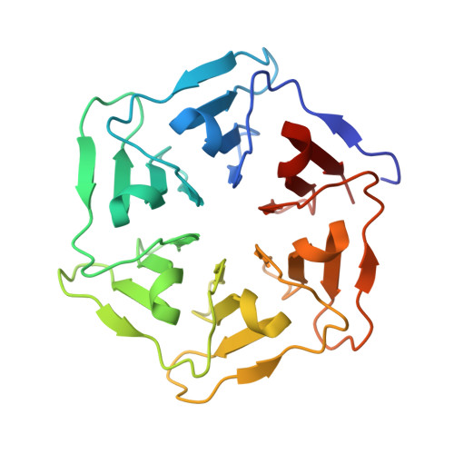
    β-propeller proteins are highly symmetrical, being composed of a repeated motif with four anti-parallel β-sheets arranged around a central axis. Recently we designed the first completely symmetrical β-propeller protein, Pizza6, consisting of six identical tandem repeats. Pizza6 is expected to prove a useful building block for bionanotechnology, and also a tool to investigate the folding and evolution of β-propeller proteins. Folding studies are made difficult by the high stability and the lack of buried Trp residues to act as monitor fluorophores, so we have designed and characterized several Trp-containing Pizza6 derivatives. In total four proteins were designed, of which three could be purified and characterized. Crystal structures confirm these mutant proteins maintain the expected structure, and a clear redshift of Trp fluorescence emission could be observed upon denaturation. Among the derivative proteins, Pizza6-AYW appears to be the most suitable model protein for future folding/unfolding kinetics studies as it has a comparable stability as natural β-propeller proteins.


  • Organizational Affiliation
    • Laboratory of Biomolecular Modelling and Design, Department of Chemistry, University of Leuven, Celestijnenlaan 200G-bus2403, Heverlee, Belgium.

Macromolecule Content 

  • Total Structure Weight: 51.72 kDa 
  • Atom Count: 4,604 
  • Modeled Residue Count: 502 
  • Deposited Residue Count: 512 
  • Unique protein chains: 1

Macromolecules

Find similar proteins by:|  3D Structure
Entity ID: 1
MoleculeChains  Sequence LengthOrganismDetailsImage
Pizza6-AYW
A, B
256synthetic constructMutation(s): 0 

Experimental Data & Validation

Experimental Data

  • Method: X-RAY DIFFRACTION
  • Resolution: 1.30 Å
  • R-Value Free:  0.192 (Depositor), 0.192 (DCC) 
  • R-Value Work:  0.155 (Depositor), 0.156 (DCC) 
  • R-Value Observed: 0.157 (Depositor) 
Space Group: P 1 21 1
Unit Cell:
Length ( Å )Angle ( ˚ )
a = 47.01α = 90
b = 70.35β = 100.6
c = 58.21γ = 90
Software Package:
Software NamePurpose
PHENIXrefinement
XDSdata reduction
Aimlessdata scaling
PHASERphasing

Structure Validation

View Full Validation Report


Entry History 

& Funding Information

Deposition Data


Funding OrganizationLocationGrant Number
FWOBelgiumG0F9316N
FWOBelgiumG051917N

Revision History  (Full details and data files)

  • Version 1.0: 2018-03-21
    Type: Initial release
  • Version 1.1: 2024-01-17
    Changes: Data collection, Database references, Refinement description