6EWZ | pdb_00006ewz

Crystal structure of RelP (SAS2) from Staphylococcus aureus bound to AMPCPP and GTP in the pre-catalytic state


Experimental Data Snapshot

  • Method: X-RAY DIFFRACTION
  • Resolution: 2.24 Å
  • R-Value Free: 
    0.234 (Depositor), 0.242 (DCC) 
  • R-Value Work: 
    0.193 (Depositor), 0.204 (DCC) 
  • R-Value Observed: 
    0.195 (Depositor) 

Starting Model: experimental
View more details

wwPDB Validation 3D Report Full Report

Validation slider image for 6EWZ

Ligand Structure Quality Assessment 


This is version 1.2 of the entry. See complete history

Literature

Structural basis for (p)ppGpp synthesis by theStaphylococcus aureussmall alarmone synthetase RelP.

Manav, M.C.Beljantseva, J.Bojer, M.S.Tenson, T.Ingmer, H.Hauryliuk, V.Brodersen, D.E.

(2018) J Biological Chem 293: 3254-3264

  • DOI: https://doi.org/10.1074/jbc.RA117.001374
  • Primary Citation Related Structures: 
    6EWZ, 6EX0

  • PubMed Abstract: 

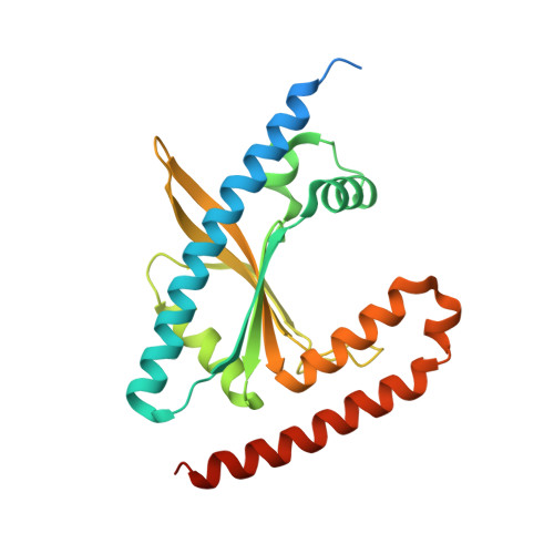
    The stringent response is a global reprogramming of bacterial physiology that renders cells more tolerant to antibiotics and induces virulence gene expression in pathogens in response to stress. This process is driven by accumulation of the intracellular alarmone guanosine-5'-di(tri)phosphate-3'-diphosphate ((p)ppGpp), which is produced by enzymes of the RelA SpoT homologue (RSH) family. The Gram-positive Firmicute pathogen, Staphylococcus aureus , encodes three RSH enzymes: a multidomain RSH (Rel) that senses amino acid starvation on the ribosome and two small alarmone synthetase (SAS) enzymes, RelQ (SAS1) and RelP (SAS2). In Bacillus subtilis , RelQ (SAS1) was shown to form a tetramer that is activated by pppGpp and inhibited by single-stranded RNA, but the structural and functional regulation of RelP (SAS2) is unexplored. Here, we present crystal structures of S. aureus RelP in two major functional states, pre-catalytic (bound to GTP and the non-hydrolyzable ATP analogue, adenosine 5'-(α,β-methylene)triphosphate (AMP-CPP)), and post-catalytic (bound to pppGpp). We observed that RelP also forms a tetramer, but unlike RelQ (SAS1), it is strongly inhibited by both pppGpp and ppGpp and is insensitive to inhibition by RNA. We also identified putative metal ion-binding sites at the subunit interfaces that were consistent with the observed activation of the enzyme by Zn 2+ ions. The structures reported here reveal the details of the catalytic mechanism of SAS enzymes and provide a molecular basis for understanding differential regulation of SAS enzymes in Firmicute bacteria.


  • Organizational Affiliation
    • From the Department of Molecular Biology and Genetics, Centre for Bacterial Stress Response and Persistence, Gustav Wieds Vej 10c, DK-8000 Aarhus C, Denmark.

Macromolecule Content 

  • Total Structure Weight: 58.35 kDa 
  • Atom Count: 3,614 
  • Modeled Residue Count: 402 
  • Deposited Residue Count: 474 
  • Unique protein chains: 1

Macromolecules

Find similar proteins by:|  3D Structure
Entity ID: 1
MoleculeChains  Sequence LengthOrganismDetailsImage
GTP pyrophosphokinase
A, B
237Staphylococcus aureusMutation(s): 0 
Gene Names: ywaCBER48_002454CEJ93_03805ERS072738_01254ERS073583_01544ERS074020_00750HMPREF3211_00175
EC: 2.7.6.5
UniProt
Find proteins for W8U368 (Staphylococcus aureus)
Explore W8U368 
Go to UniProtKB:  W8U368
Entity Groups
Sequence Clusters30% Identity50% Identity70% Identity90% Identity95% Identity100% Identity
UniProt GroupW8U368
Sequence Annotations
Expand
Reference Sequence

Small Molecules

Ligands 4 Unique
IDChains Name / Formula / InChI Key2D Diagram3D Interactions
GTP

Query on GTP


Download:Ideal Coordinates CCD File
C [auth A],
G [auth B]
GUANOSINE-5'-TRIPHOSPHATE
C10 H16 N5 O14 P3
XKMLYUALXHKNFT-UUOKFMHZSA-N
APC

Query on APC


Download:Ideal Coordinates CCD File
D [auth A],
H [auth B]
DIPHOSPHOMETHYLPHOSPHONIC ACID ADENOSYL ESTER
C11 H18 N5 O12 P3
CAWZRIXWFRFUQB-IOSLPCCCSA-N
FE

Query on FE


Download:Ideal Coordinates CCD File
F [auth A]FE (III) ION
Fe
VTLYFUHAOXGGBS-UHFFFAOYSA-N
MG

Query on MG


Download:Ideal Coordinates CCD File
E [auth A],
I [auth B]
MAGNESIUM ION
Mg
JLVVSXFLKOJNIY-UHFFFAOYSA-N

Experimental Data & Validation

Experimental Data

  • Method: X-RAY DIFFRACTION
  • Resolution: 2.24 Å
  • R-Value Free:  0.234 (Depositor), 0.242 (DCC) 
  • R-Value Work:  0.193 (Depositor), 0.204 (DCC) 
  • R-Value Observed: 0.195 (Depositor) 
Space Group: I 41 2 2
Unit Cell:
Length ( Å )Angle ( ˚ )
a = 125.12α = 90
b = 125.12β = 90
c = 219.07γ = 90
Software Package:
Software NamePurpose
PHENIXrefinement
xia2data reduction
XSCALEdata scaling
PHASERphasing

Structure Validation

View Full Validation Report


Ligand Structure Quality Assessment 


Entry History 

& Funding Information

Deposition Data


Funding OrganizationLocationGrant Number
Danish National Research FoundationDenmarkDNRF120

Revision History  (Full details and data files)

  • Version 1.0: 2018-01-24
    Type: Initial release
  • Version 1.1: 2018-03-28
    Changes: Data collection, Database references
  • Version 1.2: 2024-01-17
    Changes: Advisory, Data collection, Database references, Derived calculations, Refinement description