6EV2 | pdb_00006ev2

Crystal structure of antibody against schizophyllan in complex with laminarihexaose


Experimental Data Snapshot

  • Method: X-RAY DIFFRACTION
  • Resolution: 2.40 Å
  • R-Value Free: 
    0.233 (Depositor), 0.235 (DCC) 
  • R-Value Work: 
    0.194 (Depositor), 0.194 (DCC) 
  • R-Value Observed: 
    0.195 (Depositor) 

Starting Model: experimental
View more details

wwPDB Validation 3D Report Full Report

Validation slider image for 6EV2

Ligand Structure Quality Assessment 


This is version 2.2 of the entry. See complete history

Literature

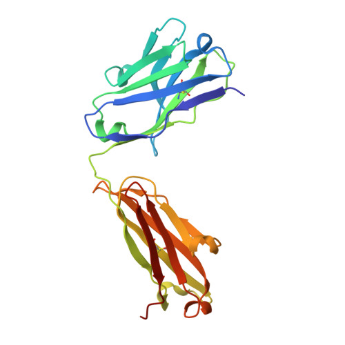
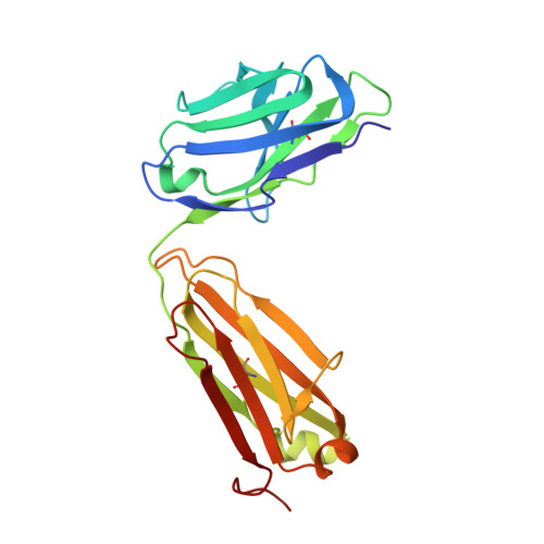
Structural insights into antigen recognition of an anti-beta-(1,6)-beta-(1,3)-D-glucan antibody.

Sung, K.H.Josewski, J.Dubel, S.Blankenfeldt, W.Rau, U.

(2018) Sci Rep 8: 13652-13652

  • DOI: https://doi.org/10.1038/s41598-018-31961-x
  • Primary Citation Related Structures: 
    6EV1, 6EV2

  • PubMed Abstract: 

    Schizophyllan (SCH) is a high molecular weight homopolysaccharide composed of a β-(1,3)-D-glucan main chain with branching β-(1,6)-bound D-glucose residues. It forms triple helices that are highly stable towards heat and extreme pH, which provides SCH with interesting properties for industrial and medical applications. The recombinant anti-SCH antibody JoJ48C11 recognizes SCH and related β-(1,6)-branched β-(1,3)-D-glucans, but details governing its specificity are not known. Here, we fill this gap by determining crystal structures of the antigen binding fragment (Fab) of JoJ48C11 in the apo form and in complex with the unbranched β-(1,3)-D-glucose hexamer laminarihexaose 3.0 and 2.4 Å resolution, respectively. Together with docking studies, this allowed construction of a JoJ48C11/triple-helical SCH complex, leading to the identification of eight amino acid residues of JoJ48C11 (Tyr27 H , His35 H , Trp47 H , Trp100 H , Asp105 H ; Asp49 L , Lys52 L , Trp90 L ) that contribute to the recognition of glucose units from all three chains of the SCH triple helix. The importance of these amino acids was confirmed by mutagenesis and ELISA-based analysis. Our work provides an explanation for the specific recognition of triple-helical β-(1,6)-branched β-(1,3)-D-glucans by JoJ48C11 and provides another structure example for anti-carbohydrate antibodies.


  • Organizational Affiliation
    • Structure and Function of Proteins, Helmholtz Centre for Infection Research, Inhoffenstraße 7, 38124, Braunschweig, Germany.

Macromolecule Content 

  • Total Structure Weight: 189.34 kDa 
  • Atom Count: 13,666 
  • Modeled Residue Count: 1,731 
  • Deposited Residue Count: 1,756 
  • Unique protein chains: 2

Macromolecules

Find similar proteins by:|  3D Structure
Entity ID: 1
MoleculeChains  Sequence LengthOrganismDetailsImage
Heavy chain
A, C, E, G
222Mus musculusMutation(s): 0 
Find similar proteins by:|  3D Structure
Entity ID: 2
MoleculeChains  Sequence LengthOrganismDetailsImage
Light chain
B, D, F, H
217Mus musculusMutation(s): 0 

Oligosaccharides

Help  
Entity ID: 3
MoleculeChains Length2D Diagram GlycosylationD Interactions
beta-D-glucopyranose-(1-3)-beta-D-glucopyranose
I
2N/A
Glycosylation Resources
GlyTouCan: G36535HU
GlyCosmos: G36535HU

Small Molecules

Ligands 1 Unique
IDChains Name / Formula / InChI Key2D Diagram3D Interactions
BGC

Query on BGC


Download:Ideal Coordinates CCD File
J [auth A]beta-D-glucopyranose
C6 H12 O6
WQZGKKKJIJFFOK-VFUOTHLCSA-N

Biologically Interesting Molecules (External Reference) 

1 Unique

Experimental Data & Validation

Experimental Data

  • Method: X-RAY DIFFRACTION
  • Resolution: 2.40 Å
  • R-Value Free:  0.233 (Depositor), 0.235 (DCC) 
  • R-Value Work:  0.194 (Depositor), 0.194 (DCC) 
  • R-Value Observed: 0.195 (Depositor) 
Space Group: P 1 21 1
Unit Cell:
Length ( Å )Angle ( ˚ )
a = 81.732α = 90
b = 131.502β = 91.5
c = 91.034γ = 90
Software Package:
Software NamePurpose
PHENIXrefinement
XDSdata reduction
autoPROCdata scaling
PHENIXphasing

Structure Validation

View Full Validation Report


Ligand Structure Quality Assessment 


Entry History 

Deposition Data

Revision History  (Full details and data files)

  • Version 1.0: 2018-09-26
    Type: Initial release
  • Version 2.0: 2020-07-29
    Type: Remediation
    Reason: Carbohydrate remediation
    Changes: Atomic model, Data collection, Derived calculations, Refinement description, Structure summary
  • Version 2.1: 2024-01-17
    Changes: Data collection, Database references, Refinement description, Structure summary
  • Version 2.2: 2024-10-23
    Changes: Structure summary