6ETM | pdb_00006etm

Atomic resolution structure of RNase A (data collection 3)


Experimental Data Snapshot

  • Method: X-RAY DIFFRACTION
  • Resolution: 0.92 Å
  • R-Value Free: 
    0.136 (Depositor), 0.122 (DCC) 
  • R-Value Work: 
    0.106 (Depositor), 0.099 (DCC) 
  • R-Value Observed: 
    0.109 (Depositor) 

wwPDB Validation 3D Report Full Report

Validation slider image for 6ETM

This is version 1.1 of the entry. See complete history

Literature

Raman-markers of X-ray radiation damage of proteins.

Vergara, A.Caterino, M.Merlino, A.

(2018) Int J Biol Macromol 111: 1194-1205

  • DOI: https://doi.org/10.1016/j.ijbiomac.2018.01.135
  • Primary Citation Related Structures: 
    6ETK, 6ETL, 6ETM, 6ETN, 6ETP, 6ETQ, 6ETR

  • PubMed Abstract: 

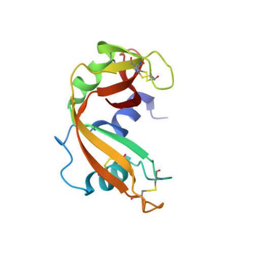
    Despite their high relevance, the mechanisms of X-ray radiation damage on protein structure yet have to be completely established. Here, we used Raman microspectrophotometry to follow X-ray-induced chemical modifications on the structure of the model protein bovine pancreatic ribonuclease (RNase A). The combination of dose-dependent Raman spectra and ultrahigh resolution (eight structures solved using data collected between 0.85 and 1.17 Å resolution on the same single crystal) allowed direct observation of several radiation damage events, including covalent bond breakages and formation of radicals. Our results are relevant for analytical photodamage detection and provide implications for a detailed understanding of the mechanisms of photoproduct formation.


  • Organizational Affiliation
    • Department of Chemical Sciences, University of Naples "Federico II", Via Cinthia, Naples I-80126, Italy; CEINGE Biotecnologie Avanzate Scarl, Via G. Salvatore, Napoli, Italy.

Macromolecule Content 

  • Total Structure Weight: 13.89 kDa 
  • Atom Count: 1,396 
  • Modeled Residue Count: 124 
  • Deposited Residue Count: 124 
  • Unique protein chains: 1

Macromolecules

Find similar proteins by:|  3D Structure
Entity ID: 1
MoleculeChains  Sequence LengthOrganismDetailsImage
Ribonuclease pancreatic124Bos taurusMutation(s): 0 
EC: 3.1.27.5 (PDB Primary Data), 4.6.1.18 (UniProt)
UniProt
Find proteins for P61823 (Bos taurus)
Explore P61823 
Go to UniProtKB:  P61823
Entity Groups
Sequence Clusters30% Identity50% Identity70% Identity90% Identity95% Identity100% Identity
UniProt GroupP61823
Sequence Annotations
Expand
Reference Sequence

Experimental Data & Validation

Experimental Data

  • Method: X-RAY DIFFRACTION
  • Resolution: 0.92 Å
  • R-Value Free:  0.136 (Depositor), 0.122 (DCC) 
  • R-Value Work:  0.106 (Depositor), 0.099 (DCC) 
  • R-Value Observed: 0.109 (Depositor) 
Space Group: P 1 21 1
Unit Cell:
Length ( Å )Angle ( ˚ )
a = 29.125α = 90
b = 37.971β = 105.83
c = 52.541γ = 90
Software Package:
Software NamePurpose
SHELXLrefinement
HKL-2000data reduction
SCALEPACKdata scaling
PHASERphasing

Structure Validation

View Full Validation Report


Entry History 

Deposition Data

Revision History  (Full details and data files)

  • Version 1.0: 2018-02-21
    Type: Initial release
  • Version 1.1: 2024-10-16
    Changes: Data collection, Database references, Structure summary