6EMH | pdb_00006emh

Crystal structure of JNK3 in complex with a pyridinylimidazole inhibitor


Experimental Data Snapshot

  • Method: X-RAY DIFFRACTION
  • Resolution: 1.76 Å
  • R-Value Free: 
    0.261 (DCC) 

Starting Model: experimental
View more details

wwPDB Validation 3D Report Full Report

Validation slider image for 6EMH

Ligand Structure Quality Assessment 


This is version 1.3 of the entry. See complete history

Literature

Structural Optimization of a Pyridinylimidazole Scaffold: Shifting the Selectivity from p38 alpha Mitogen-Activated Protein Kinase to c-Jun N-Terminal Kinase 3.

Ansideri, F.Macedo, J.T.Eitel, M.El-Gokha, A.Zinad, D.S.Scarpellini, C.Kudolo, M.Schollmeyer, D.Boeckler, F.M.Blaum, B.S.Laufer, S.A.Koch, P.

(2018) ACS Omega 3: 7809-7831

  • DOI: https://doi.org/10.1021/acsomega.8b00668
  • Primary Citation Related Structures: 
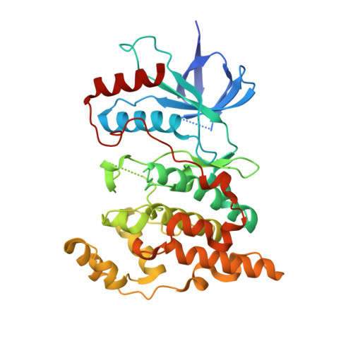
    6EKD, 6EMH, 6EQ9

  • PubMed Abstract: 

    Starting from known p38α mitogen-activated protein kinase (MAPK) inhibitors, a series of inhibitors of the c-Jun N-terminal kinase (JNK) 3 was obtained. Altering the substitution pattern of the pyridinylimidazole scaffold proved to be effective in shifting the inhibitory activity from the original target p38α MAPK to the closely related JNK3. In particular, a significant improvement for JNK3 selectivity could be achieved by addressing the hydrophobic region I with a small methyl group. Furthermore, additional structural modifications permitted to explore structure-activity relationships. The most potent inhibitor 4-(4-methyl-2-(methylthio)-1 H -imidazol-5-yl)- N -(4-morpholinophenyl)pyridin-2-amine showed an IC 50 value for the JNK3 in the low triple digit nanomolar range and its binding mode was confirmed by X-ray crystallography.


  • Organizational Affiliation
    • Department of Pharmaceutical and Medicinal Chemistry, Institute of Pharmaceutical Sciences, Eberhard Karls Universität Tübingen, Auf der Morgenstelle 8, 72076 Tübingen, Germany.

Macromolecule Content 

  • Total Structure Weight: 172.2 kDa 
  • Atom Count: 12,089 
  • Modeled Residue Count: 1,353 
  • Deposited Residue Count: 1,468 
  • Unique protein chains: 1

Macromolecules

Find similar proteins by:|  3D Structure
Entity ID: 1
MoleculeChains  Sequence LengthOrganismDetailsImage
Mitogen-activated protein kinase 10
A, B, C, D
367Homo sapiensMutation(s): 0 
Gene Names: MAPK10JNK3JNK3APRKM10SAPK1B
EC: 2.7.11.24
UniProt & NIH Common Fund Data Resources
Find proteins for P53779 (Homo sapiens)
Explore P53779 
Go to UniProtKB:  P53779
PHAROS:  P53779
GTEx:  ENSG00000109339 
Entity Groups
Sequence Clusters30% Identity50% Identity70% Identity90% Identity95% Identity100% Identity
UniProt GroupP53779
Sequence Annotations
Expand
Reference Sequence

Small Molecules

Ligands 7 Unique
IDChains Name / Formula / InChI Key2D Diagram3D Interactions
C15

Query on C15


Download:Ideal Coordinates CCD File
J [auth A],
N [auth B]
N-DODECYL-N,N-DIMETHYL-3-AMMONIO-1-PROPANESULFONATE
C17 H38 N O3 S
IZWSFJTYBVKZNK-UHFFFAOYSA-O
BGE
(Subject of Investigation/LOI)

Query on BGE


Download:Ideal Coordinates CCD File
H [auth A],
L [auth B],
Q [auth C],
V [auth D]
4-(4-methyl-1~{H}-imidazol-5-yl)-~{N}-(4-morpholin-4-ylphenyl)pyridin-2-amine
C19 H21 N5 O
KJQAULYFYNEAHK-UHFFFAOYSA-N
PEG

Query on PEG


Download:Ideal Coordinates CCD File
P [auth C],
U [auth D]
DI(HYDROXYETHYL)ETHER
C4 H10 O3
MTHSVFCYNBDYFN-UHFFFAOYSA-N
GOL

Query on GOL


Download:Ideal Coordinates CCD File
S [auth C],
Y [auth D],
Z [auth D]
GLYCEROL
C3 H8 O3
PEDCQBHIVMGVHV-UHFFFAOYSA-N
BME

Query on BME


Download:Ideal Coordinates CCD File
E [auth A],
F [auth A],
K [auth B]
BETA-MERCAPTOETHANOL
C2 H6 O S
DGVVWUTYPXICAM-UHFFFAOYSA-N
EDO

Query on EDO


Download:Ideal Coordinates CCD File
I [auth A],
M [auth B],
R [auth C],
W [auth D],
X [auth D]
1,2-ETHANEDIOL
C2 H6 O2
LYCAIKOWRPUZTN-UHFFFAOYSA-N
CL

Query on CL


Download:Ideal Coordinates CCD File
G [auth A],
O [auth C],
T [auth D]
CHLORIDE ION
Cl
VEXZGXHMUGYJMC-UHFFFAOYSA-M

Experimental Data & Validation

Experimental Data

  • Method: X-RAY DIFFRACTION
  • Resolution: 1.76 Å
  • R-Value Free:  0.261 (DCC) 
Space Group: P 21 21 21
Unit Cell:
Length ( Å )Angle ( ˚ )
a = 88.56α = 90
b = 114.26β = 90
c = 157.8γ = 90
Software Package:
Software NamePurpose
REFMACrefinement
XDSdata reduction
XSCALEdata scaling
MOLREPphasing

Structure Validation

View Full Validation Report


Ligand Structure Quality Assessment 


Entry History 

Deposition Data

Revision History  (Full details and data files)

  • Version 1.0: 2018-08-08
    Type: Initial release
  • Version 1.1: 2018-08-22
    Changes: Data collection, Database references
  • Version 1.2: 2019-10-16
    Changes: Data collection
  • Version 1.3: 2024-01-17
    Changes: Data collection, Database references, Refinement description