6EKY | pdb_00006eky

Direct-evolutioned unspecific peroxygenase from Agrocybe aegerita, in complex with 1-naphthol (II)


Experimental Data Snapshot

  • Method: X-RAY DIFFRACTION
  • Resolution: 1.23 Å
  • R-Value Free: 
    0.169 (Depositor), 0.179 (DCC) 
  • R-Value Work: 
    0.160 (Depositor), 0.170 (DCC) 
  • R-Value Observed: 
    0.161 (Depositor) 

Starting Model: experimental
View more details

wwPDB Validation 3D Report Full Report

Validation slider image for 6EKY

Ligand Structure Quality Assessment 


This is version 1.5 of the entry. See complete history

Literature

Structural Insights into the Substrate Promiscuity of a Laboratory-Evolved Peroxygenase.

Ramirez-Escudero, M.Molina-Espeja, P.Gomez de Santos, P.Hofrichter, M.Sanz-Aparicio, J.Alcalde, M.

(2018) ACS Chem Biol 13: 3259-3268

  • DOI: https://doi.org/10.1021/acschembio.8b00500
  • Primary Citation Related Structures: 
    5OXT, 5OXU, 5OY1, 5OY2, 6EKW, 6EKX, 6EKY, 6EKZ, 6EL0, 6EL4

  • PubMed Abstract: 

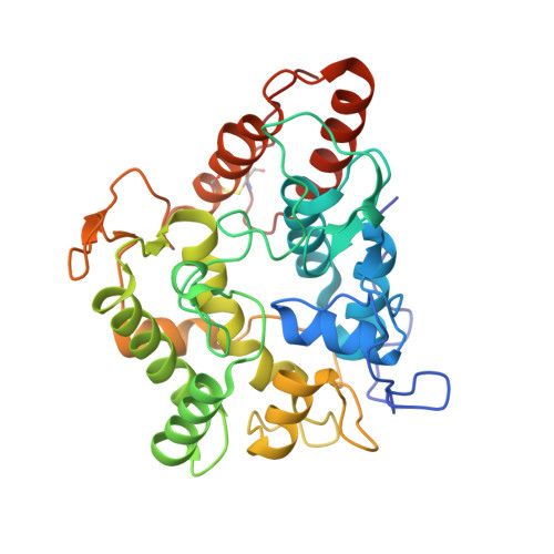
    Because of their minimal requirements, substrate promiscuity and product selectivity, fungal peroxygenases are now considered to be the jewel in the crown of C-H oxyfunctionalization biocatalysts. In this work, the crystal structure of the first laboratory-evolved peroxygenase expressed by yeast was determined at a resolution of 1.5 Å. Notable differences were detected between the evolved and native peroxygenase from Agrocybe aegerita, including the presence of a full N-terminus and a broader heme access channel due to the mutations that accumulated through directed evolution. Further mutagenesis and soaking experiments with a palette of peroxygenative and peroxidative substrates suggested dynamic trafficking through the heme channel as the main driving force for the exceptional substrate promiscuity of peroxygenase. Accordingly, this study provides the first structural evidence at an atomic level regarding the mode of substrate binding for this versatile biocatalyst, which is discussed within a biological and chemical context.


  • Organizational Affiliation
    • Department of Crystallography & Structural Biology, Institute of Physical Chemistry "Rocasolano" , CSIC , 28006 Madrid , Spain.

Macromolecule Content 

  • Total Structure Weight: 39.36 kDa 
  • Atom Count: 3,231 
  • Modeled Residue Count: 324 
  • Deposited Residue Count: 328 
  • Unique protein chains: 1

Macromolecules

Find similar proteins by:|  3D Structure
Entity ID: 1
MoleculeChains  Sequence LengthOrganismDetailsImage
Aromatic peroxygenase328Cyclocybe aegeritaMutation(s): 4 
Gene Names: APO1
EC: 1.11.2.1
UniProt
Find proteins for B9W4V6 (Cyclocybe aegerita)
Explore B9W4V6 
Go to UniProtKB:  B9W4V6
Entity Groups
Sequence Clusters30% Identity50% Identity70% Identity90% Identity95% Identity100% Identity
UniProt GroupB9W4V6
Glycosylation
Glycosylation Sites: 5
Sequence Annotations
Expand
Reference Sequence

Small Molecules

Ligands 8 Unique
IDChains Name / Formula / InChI Key2D Diagram3D Interactions
HEM

Query on HEM


Download:Ideal Coordinates CCD File
B [auth A]PROTOPORPHYRIN IX CONTAINING FE
C34 H32 Fe N4 O4
KABFMIBPWCXCRK-RGGAHWMASA-L
NAG

Query on NAG


Download:Ideal Coordinates CCD File
E [auth A],
F [auth A],
G [auth A],
H [auth A],
I [auth A]
2-acetamido-2-deoxy-beta-D-glucopyranose
C8 H15 N O6
OVRNDRQMDRJTHS-FMDGEEDCSA-N
1NP

Query on 1NP


Download:Ideal Coordinates CCD File
N [auth A],
O [auth A],
P [auth A],
Q [auth A],
R [auth A]
1-NAPHTHOL
C10 H8 O
KJCVRFUGPWSIIH-UHFFFAOYSA-N
PO4

Query on PO4


Download:Ideal Coordinates CCD File
J [auth A]PHOSPHATE ION
O4 P
NBIIXXVUZAFLBC-UHFFFAOYSA-K
GOL

Query on GOL


Download:Ideal Coordinates CCD File
GA [auth A],
K [auth A],
L [auth A],
M [auth A]
GLYCEROL
C3 H8 O3
PEDCQBHIVMGVHV-UHFFFAOYSA-N
CL

Query on CL


Download:Ideal Coordinates CCD File
D [auth A]CHLORIDE ION
Cl
VEXZGXHMUGYJMC-UHFFFAOYSA-M
MOH

Query on MOH


Download:Ideal Coordinates CCD File
AA [auth A]
BA [auth A]
CA [auth A]
DA [auth A]
EA [auth A]
AA [auth A],
BA [auth A],
CA [auth A],
DA [auth A],
EA [auth A],
FA [auth A],
S [auth A],
T [auth A],
U [auth A],
V [auth A],
W [auth A],
X [auth A],
Y [auth A],
Z [auth A]
METHANOL
C H4 O
OKKJLVBELUTLKV-UHFFFAOYSA-N
MG

Query on MG


Download:Ideal Coordinates CCD File
C [auth A]MAGNESIUM ION
Mg
JLVVSXFLKOJNIY-UHFFFAOYSA-N

Experimental Data & Validation

Experimental Data

  • Method: X-RAY DIFFRACTION
  • Resolution: 1.23 Å
  • R-Value Free:  0.169 (Depositor), 0.179 (DCC) 
  • R-Value Work:  0.160 (Depositor), 0.170 (DCC) 
  • R-Value Observed: 0.161 (Depositor) 
Space Group: P 1 21 1
Unit Cell:
Length ( Å )Angle ( ˚ )
a = 51.18α = 90
b = 57.969β = 110.13
c = 60.975γ = 90
Software Package:
Software NamePurpose
REFMACrefinement
XDSdata reduction
Aimlessdata scaling
MOLREPphasing

Structure Validation

View Full Validation Report


Ligand Structure Quality Assessment 


Entry History 

Deposition Data

Revision History  (Full details and data files)

  • Version 1.0: 2018-12-12
    Type: Initial release
  • Version 1.1: 2018-12-19
    Changes: Data collection, Database references
  • Version 1.2: 2019-04-03
    Changes: Data collection, Database references
  • Version 1.3: 2020-07-29
    Type: Remediation
    Reason: Carbohydrate remediation
    Changes: Data collection, Derived calculations, Structure summary
  • Version 1.4: 2024-01-17
    Changes: Data collection, Database references, Refinement description, Structure summary
  • Version 1.5: 2024-10-16
    Changes: Structure summary