6EK8 | pdb_00006ek8

YaxB from Yersinia enterocolitica


Experimental Data Snapshot

  • Method: X-RAY DIFFRACTION
  • Resolution: 4.00 Å
  • R-Value Free: 
    0.339 (Depositor), 0.336 (DCC) 
  • R-Value Work: 
    0.326 (Depositor), 0.320 (DCC) 
  • R-Value Observed: 
    0.326 (Depositor) 

Starting Model: experimental
View more details

wwPDB Validation 3D Report Full Report

Validation slider image for 6EK8

This is version 1.1 of the entry. See complete history

Literature

Structure and mechanism of the two-component alpha-helical pore-forming toxin YaxAB.

Brauning, B.Bertosin, E.Praetorius, F.Ihling, C.Schatt, A.Adler, A.Richter, K.Sinz, A.Dietz, H.Groll, M.

(2018) Nat Commun 9: 1806-1806

  • DOI: https://doi.org/10.1038/s41467-018-04139-2
  • Primary Citation Related Structures: 
    6EK4, 6EK7, 6EK8, 6EL1

  • PubMed Abstract: 

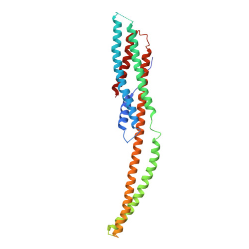
    Pore-forming toxins (PFT) are virulence factors that transform from soluble to membrane-bound states. The Yersinia YaxAB system represents a family of binary α-PFTs with orthologues in human, insect, and plant pathogens, with unknown structures. YaxAB was shown to be cytotoxic and likely involved in pathogenesis, though the molecular basis for its two-component lytic mechanism remains elusive. Here, we present crystal structures of YaxA and YaxB, together with a cryo-electron microscopy map of the YaxAB complex. Our structures reveal a pore predominantly composed of decamers of YaxA-YaxB heterodimers. Both subunits bear membrane-active moieties, but only YaxA is capable of binding to membranes by itself. YaxB can subsequently be recruited to membrane-associated YaxA and induced to present its lytic transmembrane helices. Pore formation can progress by further oligomerization of YaxA-YaxB dimers. Our results allow for a comparison between pore assemblies belonging to the wider ClyA-like family of α-PFTs, highlighting diverse pore architectures.


  • Organizational Affiliation
    • Center for Integrated Protein Science Munich (CIPSM), Department of Chemistry, Chair of Biochemistry, Technische Universität München, Lichtenbergstrasse 4, 85747, Garching, Germany. bastian.braeuning@tum.de.

Macromolecule Content 

  • Total Structure Weight: 39.25 kDa 
  • Atom Count: 2,002 
  • Modeled Residue Count: 249 
  • Deposited Residue Count: 344 
  • Unique protein chains: 1

Macromolecules

Find similar proteins by:|  3D Structure
Entity ID: 1
MoleculeChains  Sequence LengthOrganismDetailsImage
YaxB344Yersinia enterocoliticaMutation(s): 0 
Gene Names: YE1985
UniProt
Find proteins for A1JM52 (Yersinia enterocolitica serotype O:8 / biotype 1B (strain NCTC 13174 / 8081))
Explore A1JM52 
Go to UniProtKB:  A1JM52
Entity Groups
Sequence Clusters30% Identity50% Identity70% Identity90% Identity95% Identity100% Identity
UniProt GroupA1JM52
Sequence Annotations
Expand
Reference Sequence

Experimental Data & Validation

Experimental Data

  • Method: X-RAY DIFFRACTION
  • Resolution: 4.00 Å
  • R-Value Free:  0.339 (Depositor), 0.336 (DCC) 
  • R-Value Work:  0.326 (Depositor), 0.320 (DCC) 
  • R-Value Observed: 0.326 (Depositor) 
Space Group: P 65 2 2
Unit Cell:
Length ( Å )Angle ( ˚ )
a = 111.7α = 90
b = 111.7β = 90
c = 169.66γ = 120
Software Package:
Software NamePurpose
REFMACrefinement
XDSdata reduction
XSCALEdata scaling
PHASERphasing

Structure Validation

View Full Validation Report


Entry History 

Deposition Data

Revision History  (Full details and data files)

  • Version 1.0: 2018-05-16
    Type: Initial release
  • Version 1.1: 2024-01-17
    Changes: Data collection, Database references, Refinement description