6EJG | pdb_00006ejg

CRYSTAL STRUCTURE OF HUMAN CD81 LARGE EXTRACELLULAR LOOP IN COMPLEX WITH SINGLE CHAIN FV FRAGMENT 4


Experimental Data Snapshot

  • Method: X-RAY DIFFRACTION
  • Resolution: 2.82 Å
  • R-Value Free: 
    0.263 (Depositor), 0.267 (DCC) 
  • R-Value Work: 
    0.227 (Depositor), 0.232 (DCC) 
  • R-Value Observed: 
    0.229 (Depositor) 

wwPDB Validation 3D Report Full Report

Validation slider image for 6EJG

This is version 1.2 of the entry. See complete history

Literature

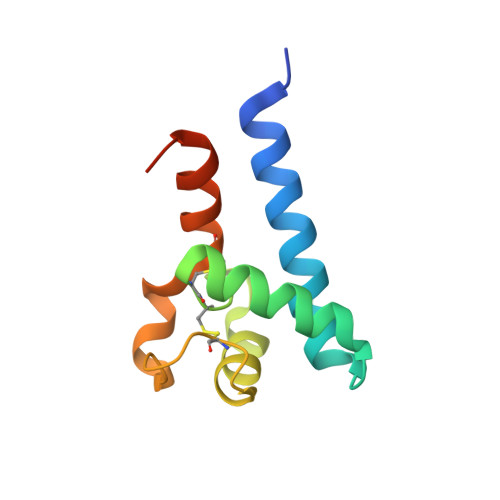
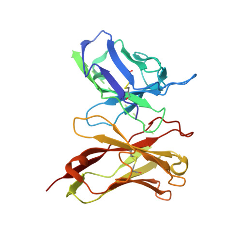
Structure-Guided Combinatorial Engineering Facilitates Affinity and Specificity Optimization of Anti-CD81 Antibodies.

Nelson, B.Adams, J.Kuglstatter, A.Li, Z.Harris, S.F.Liu, Y.Bohini, S.Ma, H.Klumpp, K.Gao, J.Sidhu, S.S.

(2018) J Mol Biology 430: 2139-2152

  • DOI: https://doi.org/10.1016/j.jmb.2018.05.018
  • Primary Citation Related Structures: 
    6EJG, 6EJM, 6EK2

  • PubMed Abstract: 

    Hepatitis C viral infection is the major cause of chronic hepatitis that affects as many as 71 million people worldwide. Rather than target the rapidly shifting viruses and their numerous serotypes, four independent antibodies were made to target the host antigen CD81 and were shown to block hepatitis C viral entry. The single-chain variable fragment of each antibody was crystallized in complex with the CD81 large extracellular loop in order to guide affinity maturation of two distinct antibodies by phage display. Affinity maturation of antibodies using phage display has proven to be critical to therapeutic antibody development and typically involves modification of the paratope for increased affinity, improved specificity, enhanced stability or a combination of these traits. One antibody was engineered for increased affinity for human CD81 large extracellular loop that equated to increased efficacy, while the second antibody was engineered for cross-reactivity with cynomolgus CD81 to facilitate animal model testing. The use of structures to guide affinity maturation library design demonstrates the utility of combining structural analysis with phage display technologies.


  • Organizational Affiliation
    • Banting and Best Department of Medical Research and Department of Medical Genetics, The Donnelly Centre for Cellular and Biomolecular Research, University of Toronto, 160 College Street, Toronto, ON M5S 3E1, Canada.

Macromolecule Content 

  • Total Structure Weight: 74.64 kDa 
  • Atom Count: 4,971 
  • Modeled Residue Count: 640 
  • Deposited Residue Count: 700 
  • Unique protein chains: 2

Macromolecules

Find similar proteins by:|  3D Structure
Entity ID: 1
MoleculeChains  Sequence LengthOrganismDetailsImage
CD81 antigen
A, B
99Homo sapiensMutation(s): 0 
Gene Names: CD81TAPA1TSPAN28
UniProt & NIH Common Fund Data Resources
Find proteins for P60033 (Homo sapiens)
Explore P60033 
Go to UniProtKB:  P60033
PHAROS:  P60033
GTEx:  ENSG00000110651 
Entity Groups
Sequence Clusters30% Identity50% Identity70% Identity90% Identity95% Identity100% Identity
UniProt GroupP60033
Sequence Annotations
Expand
Reference Sequence
Find similar proteins by:|  3D Structure
Entity ID: 2
MoleculeChains  Sequence LengthOrganismDetailsImage
SINGLE CHAIN FV FRAGMENT
C, D
251Mus musculusMutation(s): 0 

Experimental Data & Validation

Experimental Data

  • Method: X-RAY DIFFRACTION
  • Resolution: 2.82 Å
  • R-Value Free:  0.263 (Depositor), 0.267 (DCC) 
  • R-Value Work:  0.227 (Depositor), 0.232 (DCC) 
  • R-Value Observed: 0.229 (Depositor) 
Space Group: C 2 2 21
Unit Cell:
Length ( Å )Angle ( ˚ )
a = 78.635α = 90
b = 170.905β = 90
c = 185.96γ = 90
Software Package:
Software NamePurpose
REFMACrefinement
SCALEPACKdata scaling
DENZOdata reduction
PHASERphasing

Structure Validation

View Full Validation Report


Entry History 

Deposition Data

Revision History  (Full details and data files)

  • Version 1.0: 2018-05-30
    Type: Initial release
  • Version 1.1: 2018-06-20
    Changes: Data collection, Database references
  • Version 1.2: 2024-10-23
    Changes: Data collection, Database references, Structure summary