


Crystal structure of the kinase domain of the Q207E mutant of ACVR1 (ALK2) in complex with K02288
Williams, E.P., Canning, P., Sanvitale, C.E., Bullock, A.N.To be published.
Experimental Data Snapshot
Starting Model: experimental
View more details
Entity ID: 1 | |||||
|---|---|---|---|---|---|
| Molecule | Chains | Sequence Length | Organism | Details | Image |
| Activin receptor type-1 | 340 | Homo sapiens | Mutation(s): 1 Gene Names: ACVR1, ACVRLK2 EC: 2.7.11.30 | 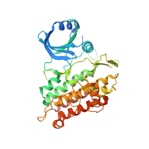 | |
UniProt & NIH Common Fund Data Resources | |||||
PHAROS: Q04771 GTEx: ENSG00000115170 | |||||
Entity Groups | |||||
| Sequence Clusters | 30% Identity50% Identity70% Identity90% Identity95% Identity100% Identity | ||||
| UniProt Group | Q04771 | ||||
Sequence AnnotationsExpand | |||||
Reference Sequence | |||||
| Ligands 2 Unique | |||||
|---|---|---|---|---|---|
| ID | Chains | Name / Formula / InChI Key | 2D Diagram | 3D Interactions | |
| A3F (Subject of Investigation/LOI) Download:Ideal Coordinates CCD File | B [auth A] | 3-[6-amino-5-(3,4,5-trimethoxyphenyl)pyridin-3-yl]phenol C20 H20 N2 O4 CJLMANFTWLNAKC-UHFFFAOYSA-N |  | ||
| EDO Download:Ideal Coordinates CCD File | C [auth A] D [auth A] E [auth A] F [auth A] G [auth A] | 1,2-ETHANEDIOL C2 H6 O2 LYCAIKOWRPUZTN-UHFFFAOYSA-N |  | ||
| Length ( Å ) | Angle ( ˚ ) |
|---|---|
| a = 81.56 | α = 90 |
| b = 66.99 | β = 91.04 |
| c = 62.22 | γ = 90 |
| Software Name | Purpose |
|---|---|
| REFMAC | refinement |
| iMOSFLM | data reduction |
| Aimless | data scaling |
| PHASER | phasing |


